Working with the Core Facilities
Our Core Facilities serve as key technical and knowledge hubs, providing access to world-leading facilities for the Cambridge cancer research community.
With their cutting-edge equipment and technical expertise, they can support research teams to adopt new techniques and create new approaches to further the cancer research field and improve outcomes for patients.
Researchers from outside of the Institute can request to work with our Core Facilities on a fee-for-service basis. We can support a wide variety of research, although due to capacity we prioritise supporting cancer-related projects. If you are interested in working with us, please use the contact form below.
Equipment and capabilities
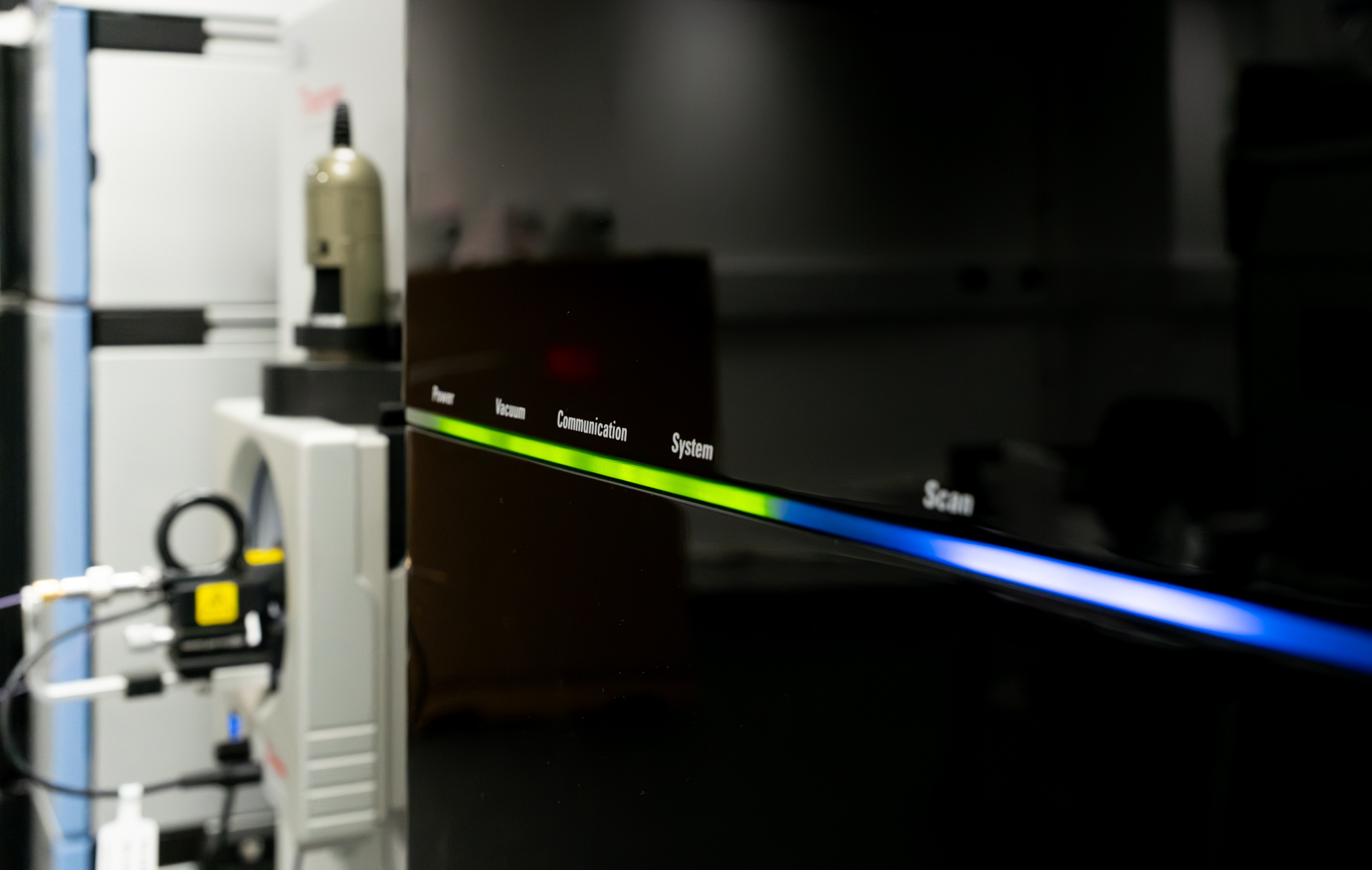
Bioanalytical mass spectrometry
The Bioanalytical mass spectrometry core facility can currently provide the following services and equipment:
- GCP regulated bioanalysis
- Drug quantitation in Clinical Trials (primary & secondary endpoints)
- Non-regulated drug quantitation
- Preclinical research
- Measurement of small molecule biomarkers, for example fumarate/malate
- Semi-targeted metabolomic screens
- TCA cycle, pentose phosphate pathway, etc.
- In vivo Drug Discovery service
- Pharmacokinetics (PK)
- Maximum tolerated dose
- Efficacy studies
Bioinformatics and statistical data analysis
The Bioinformatics Core Facility has considerable experience in analysing datasets generated by high-throughput technologies and can support research projects that employ a range of experimental approaches.
We can provide analysis support for:
- RNA-seq for differential gene expression between sample groups, e.g. treated vs. untreated cell lines, and single cell RNA-seq for identification and characterisation of sub-populations of cells
- ChIP-seq to investigate how transcriptional regulation is altered in cancers by locating binding sites of regulatory proteins and determining which of these are differentially bound under different conditions
- Whole genome, exome and amplicon sequencing to explore variation in cancer genomes, identifying single nucleotide variants, small insertions/deletions, copy number aberration and genomic rearrangements
- Quantitative proteomics including the analysis of tandem mass tag (TMT) mass spectrometry data to look at changes in protein abundance following treatment or a perturbation
- Statistical analysis of a wide range of data types including application of mixed models for tumour growth curve data and survival analysis comparing treatment groups or disease subtypes
Flow and Mass Cytometry
The Flow and Mass Cytometry Core Facility can provide the following equipment and services for use by researchers based outside of the Institute:
- Regular and spectral flow cytometer analysers available to use independently or assisted
- ImageStream, Imaging flow cytometer – for fluorescence localisation and morphology
- CyTOF mass cytometer – high parameter analysis service
- CyTOF imaging mass cytometer – high parameter tissue imaging service
- Cell sorting service, into tubes or plates
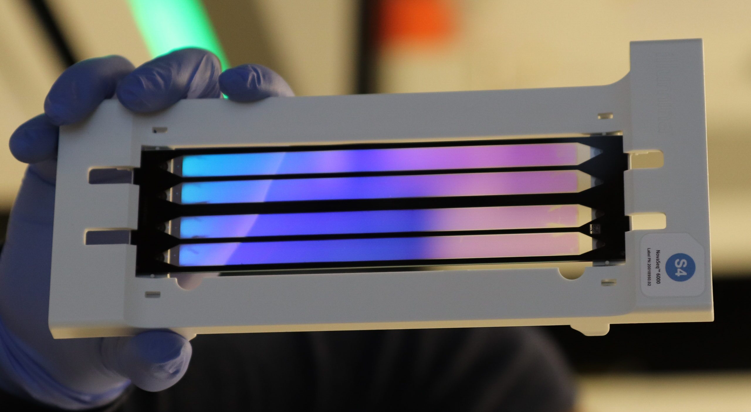
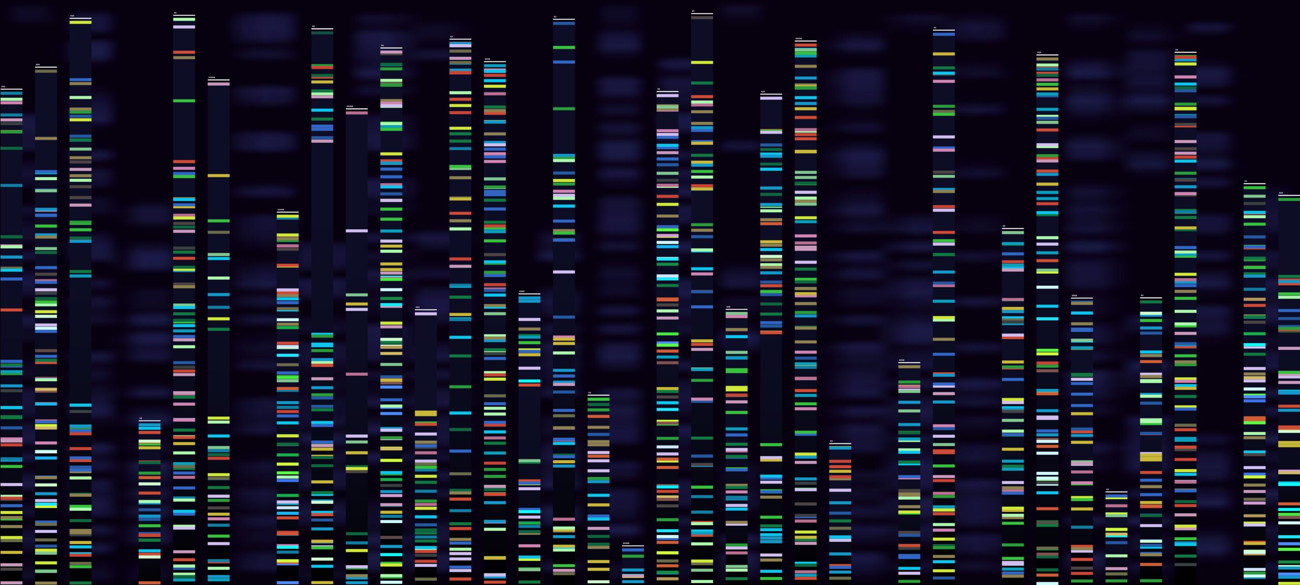
Genomics
The Genomics Core Facility can provide the following services:
- Illumina sequencing using the next-generation sequencing technologies (Illumina NovaSeqX Plus, Illumina NovaSeq6000, Illumina NextSeq2000, Illumina Miseq). Long read sequencing using ONT PromethION24 (Nanopore). These allow unbiased genome-wide experiments to be performed that enable researchers to see, at base-pair resolution, what the underlying sequence differences are in cancer genomes.
- Library Preparation services; typically for genomes, exomes, transcriptomes and ChIP-seq experiments. Our workflows include low and high DNA inputs so as low and high nucleic acids quality (like FFPE samples).
Both of the above services are available to be completed by our core members, or as a self-service option (once training is complete).
- Single cell library preparation service, available using 10X Genomics technology (all types of workflow including spatial Visium platform or plate-based methods using a low volume liquid handling robot (SPT Labtech Mosquito HV).
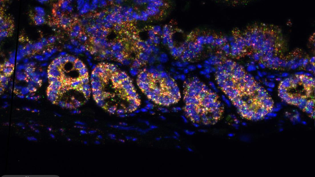
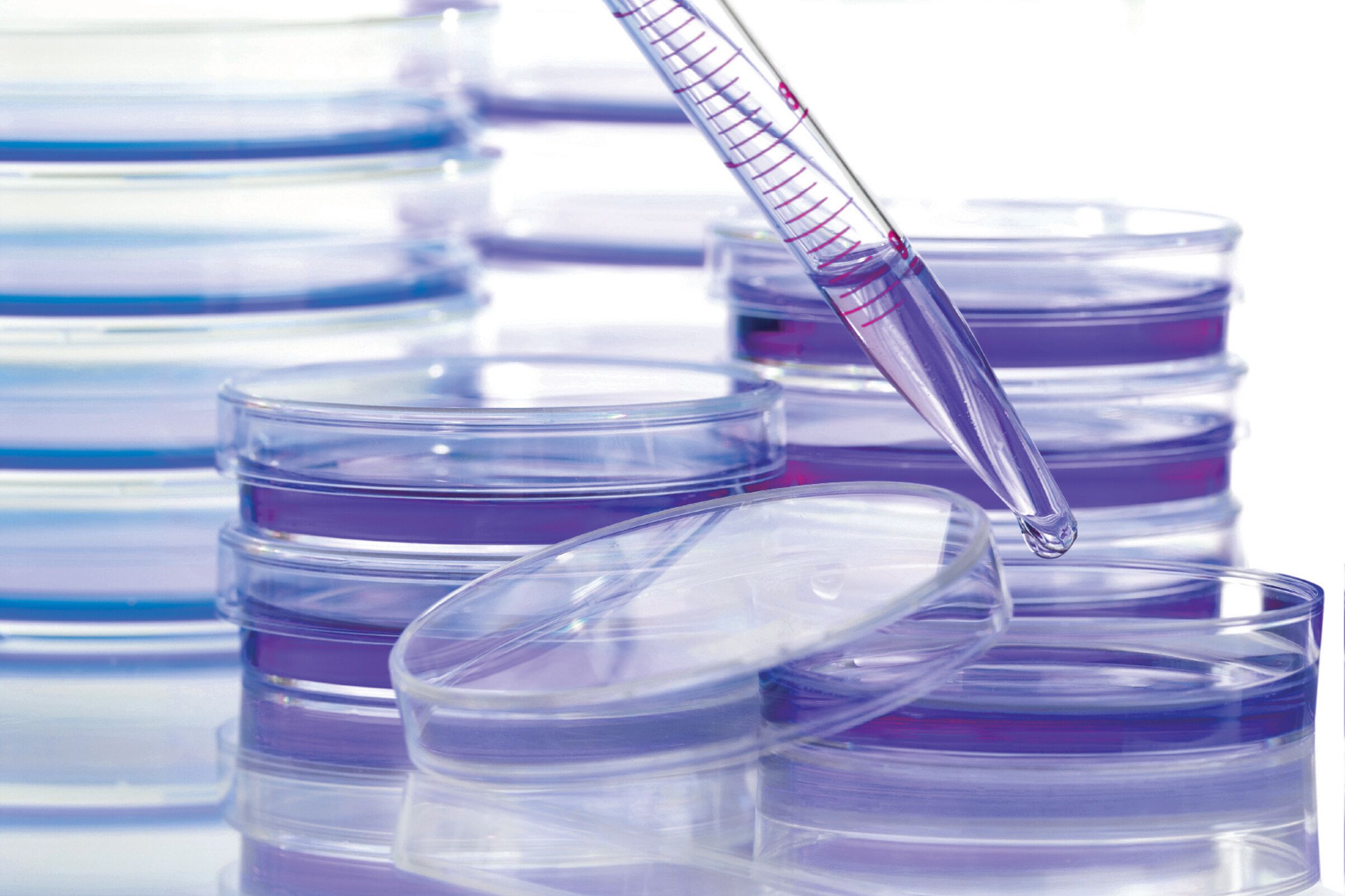
Histopathology
The Histopathology Core Facility can currently provide the following services, equipment and associated training:
Services
- Routine Immunohistochemistry (IHC) – where antibodies have already been validated on the Leica Bond
- RNAscope In situ hybridisation (ISH) – automated on the Leica Bond
- Brightfield Scanning (Leica Aperio)
This work is charged per slide.
Equipment
We can provide training and access to:
- Laser Capture Microdissector (isolate specific cells of interest ready for downstream applications)
- Vibratome (allows sectioning without the need to freeze or embed)
Use of our equipment, and the required training to do so, is charged at an hourly rate.
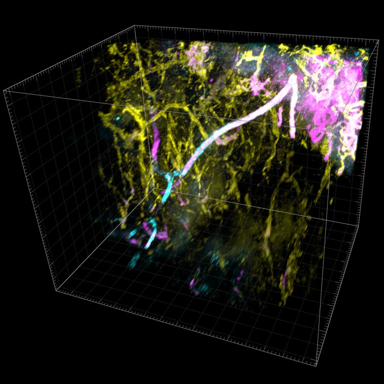
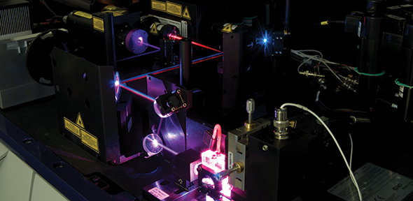
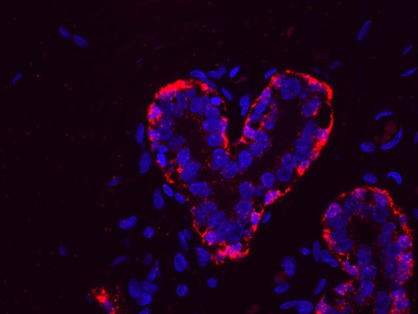
Light Microscopy
The Microscopy Core Facility can currently provide the following services and equipment:
- Advanced live-cell imaging
- STED super-resolution microscopy (Leica SP8 STED)
- Quantitative high content image acquisition (Revvity Operetta and Opera Phenix Plus)
- Light sheet microscopy of living and cleared samples
Pre-clinical Genome Editing
The Pre-clinical Genome Editing Core Facility can support researchers outside of the Institute with the following:
- CRISPR and Functional Genomics, including CRISPR engineering of cell lines, organoids and more complex systems; single gene <> sub-genome scale CRISPR editing (we don’t do genome-wide CRISPR libraries); and broad experience of CRISPR tech (KO, KI, Prime editing, Base editing, inhibition, etc.)
- Transgenics such as CRISPR engineering of embryos and early development (no live animals produced)
- Mouse model generation (for external researchers with a footprint in our Biological Resource Unit)
- Pre-clinical Therapeutic Studies (must have a PPL and cage space within the BRU) such as xenografting and multi-agent chemotherapeutics and radiotherapy.
Proteomics
The Proteomics Core Facility specialises in a suite of proteomic analyses that include but not limited to:
- Ultra deep quantitative proteome analysis
- Quantitative analysis of post-translationally modified peptides including: phosphorylated, acetylated and ubiquitinylated peptides
- Targeted quantification of peptides and proteins
- Cross-linking mass spectometry (XL-MS)
- Chromatin proteomics (protein composition & dynamics)
- Proteomic analysis of clinical tissue sections (FFPE & frozen)
- Specialised proteomic analysis of biofluids, including plasma, using our in-house Seer platform
- Secretome analysis
Research Instrumentation and Cell Services
The RICS Core Facility is able to provide access to a wide range of laboratory equipment and cell services:
Equipment
- Plate readers and spectrophotometers (fluorescence, absorbance and luminescence)
- Imagers and scanners (fluorescent, visible and radioactive samples)
- Seahorse XFe96 (cell metabolism analysis)
- Molecular Biology equipment (nucleic acid extraction, quantification and PCR/qPCR)
- Automated Western blotting
Cell Services (quality control testing)
- Mycoplasma testing
- Human and mouse cell line authentication
- Sterility testing
Get in touch
Fields marked with an asterisk * are mandatory.
"*" indicates required fields