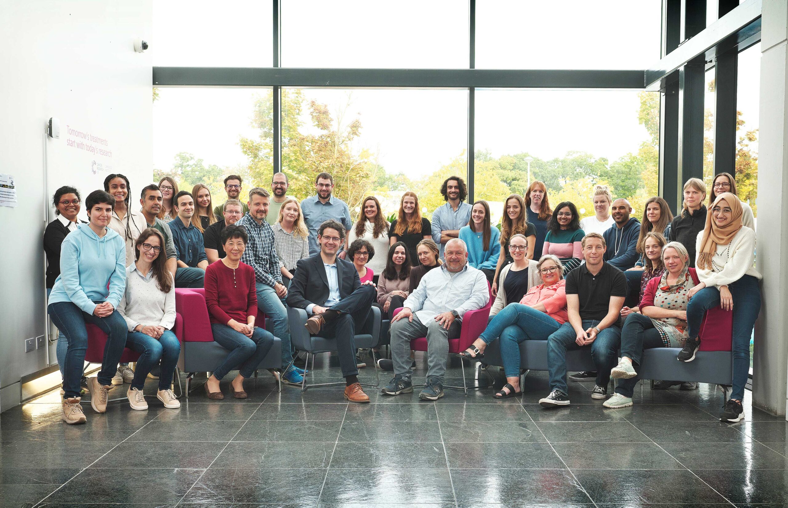
Hannon Group
Small RNAs and mammalian genomics
Introduction
The Hannon Group has a long-standing interest in small RNAs, having defined many key components of the RNAi and microRNA pathways (Hammond et al., 2000, Bernstein et al., 2001, Hammon et al., 2001 and Liu et al., 2004). This led to a particular interest in a germline-specific class of small RNAs, called PIWI-interacting RNAs (piRNAs), which form part of a sophisticated innate immune system that essentially distinguish genes (self) from transposons (nonself) and selectively silences potentially active mobile elements (Girard et al., 2006, Brennecke et al., 2007, 2008, Malone et al., 2009).
Over the past decade, we have uncovered the components of the piRNA pathway and continue to uncover exciting new discoveries of the host-parasite arms race (Czech et al. 2013, Muerdter et al 2013, Czech et al., 2018, Munafo et al., 2019, Fabry et al., 2019, Kneuss et al., 2019, Eastwood et al., 2021, Munafo et al., 2021, Fabry et al., 2021, van Lopik et al., 2023). Our interest in small RNAs and RNAi motivated the development of short hairpin (shRNAs) as powerful tools for mammalian genetics (Paddison et al., 2002). These advances along with our development of in-situ oligonucleotide synthesis on microarrays as a way to generate complex oligonucleotide libraries (Cleary et al., 2004), allowed the development of genome-wide shRNA libraries for several animal models, which are now publicly available and widely used (Silva et al., 2008). These oligonucleotide technologies also facilitated the development of exome capture (Hodges et al., 2007), and related genome partitioning strategies, now widely utilised in cancer genomics, as well as the development of CRISPR libraires.
More recently, the group has pioneered dual guide CRISPR screening approaches, defining rules that make potent guide pairs to maximise loss of function outcomes (Erard et al., 2017).
Current research within the group involves a continued interest in small RNAs and chromatin biology, understanding the biology of early breast cancer, and developing tools and techniques to track tumour heterogeneity.

Professor Greg Hannon
Senior Group Leader
Research themes
Small RNAs and chromatin biology
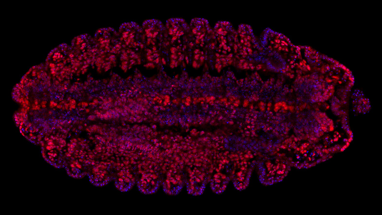
Transposons form substantial fractions of most eukaryotic genomes. Their potentially deleterious impact on host fitness has driven the evolution of transposon control mechanisms, whose function is critical in animal germ cells. The inability to silence even a single element in Drosophila is sufficient to cause complete sterility. However, the relationship between transposons and their hosts is complex. Transposons have been co-opted to provide critical regulatory elements during development and underlie telomere maintenance in numerous species.
The piRNA pathway is a critical transposon control mechanism, vital for germ cell integrity and fertility throughout the animal kingdom. Though we have a general outline of how the pathway functions, we still have a tremendous amount to learn about many aspects of piRNA biology. We use Drosophila as a model system to deepen our mechanistic understanding of piRNA biology, drawing upon genetics, biochemistry, molecular and structural biology, high resolution and live-cell imaging, as well as computational and evolutionary biology.
As part of this research programme, we seek to understand (1) how genomic loci are defined as sources of piRNAs and how their products are fated for processing, (2) how the organization of the piRNA biogenesis machinery on the surface of mitochondria enables processing of precursor transcripts, (3) how Piwi-piRNA complexes direct co-transcriptional silencing of target loci, and (4) whether the piRNA pathway shows spatio-temporal variation in its function during different stages of development.
Early breast cancer
Ductal carcinoma in situ (DCIS) is often referred to as early-stage breast cancer. These abnormal cells are localized to the milk ducts, and as they are technically still external to the body, they are not considered an invasive cancer. However, DCIS can progress to invasive ductal carcinoma (IDC), where cells have broken out of the duct and begun to invade the breast tissue, developing into one of many subtypes of breast cancer. One of the major challenges that we are addressing is identifying what causes some women to develop IDC whereas others do not, and how best to treat women diagnosed with DCIS to avoid over treatment (Rebbeck et al., 2022). We are using a combination of laser capture microscopy, to isolate small, specific areas of breast tissue with low input RNA-seq to look for shared and distinct traits in this disease.
Aside from well-known genetic factors, what determines susceptibility to breast cancer development is still an unknown. Using a combination of approaches involving imaging, low input RNA sequencing, lineage tracing and genetic manipulation we are investigating the very early initiating events and processes that occur in response to damage and how this may differ in cells that are more, or less, vulnerable to oncogenic transformation. We are using the protective benefits of pregnancy, an evolutionary conserved phenomenon, to understand these very early initiation events with the goal to find new dependencies to target.
Tumour heterogeneity
Motivated by the importance of tumour heterogeneity to patient outcomes, but a lack of models which functionally interrogate the underlying biology, we have developed strategies to study heterogeneity in animal models of cancer.
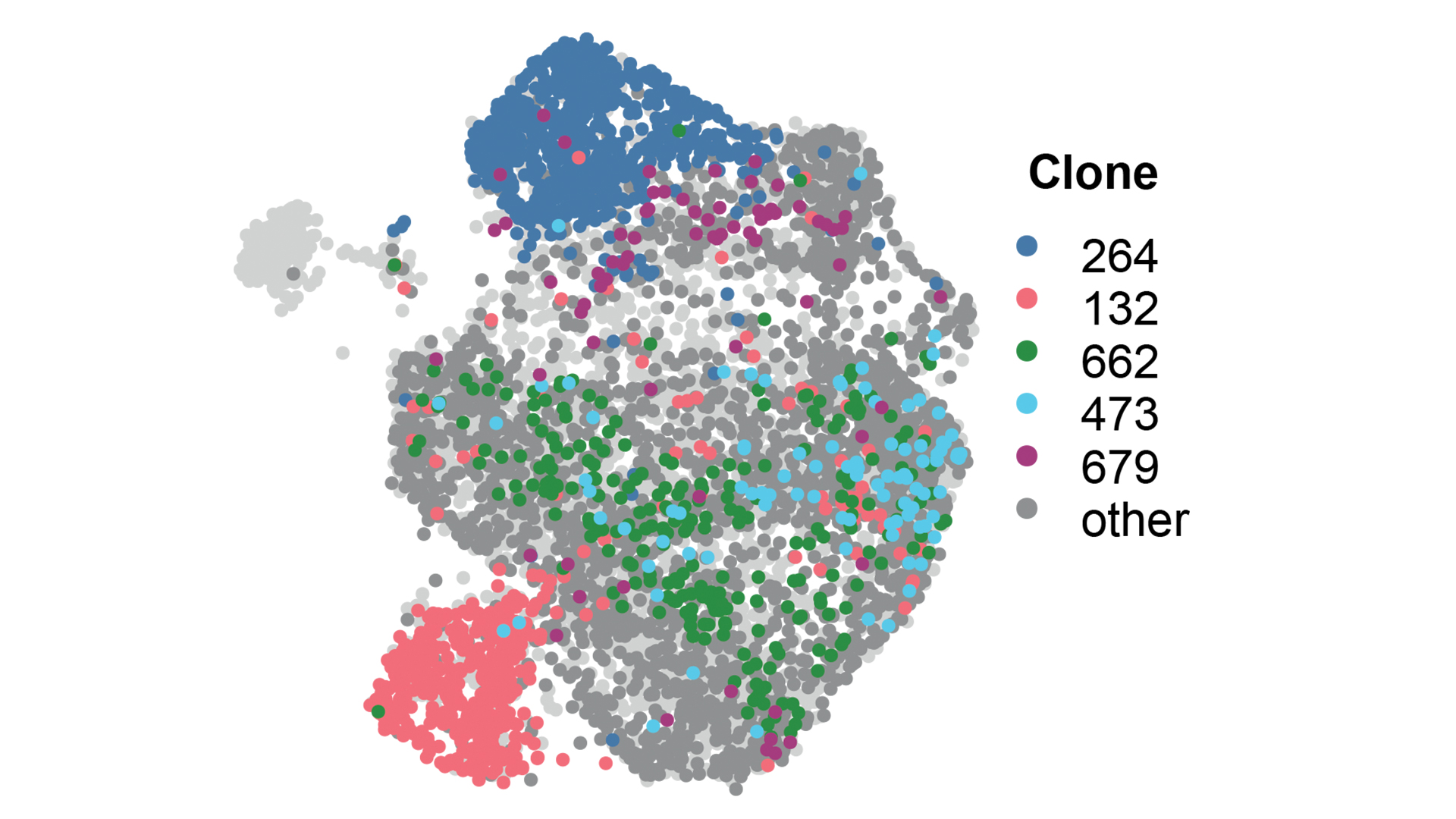
The initial studies using DNA barcoding strategies uncovered unexpected roles for vasculogenic mimicry (VM) (Wagenblast et al., 2015) and asparagine bioavailability (Knott et al., 2018) in different stages of metastasis. These observations also inspired the development of WILD-seq to determine how tumour heterogeneity impacts therapeutic response in vivo using adaptations to our barcoding approach that facilitate simultaneous tracking of clonal lineage and gene expression using single cell RNA-sequencing (Wild et al., 2022). A major finding of this work was the identification of a metabolic vulnerability of chemotherapy resistant cells, which can be targeted therapeutically.
Ongoing work in the lab seeks to understand the role of metabolism more broadly in drug resistance and tumour progression. We are also expanding the reach of WILD-seq to diverse tumour types and model systems, including patient-derived cell and xenograft models, as well as adding new capabilities on top of lineage tracing and transcriptomics.
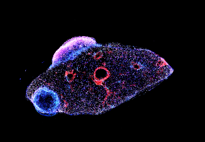
One functional output of tumour heterogeneity that we continue to study is vasculogenic mimicry (VM), a process by which tumours form pseudo blood vessels lined with tumour cells that have acquired endothelial-like properties. Building on our prior observations of VM driving metastasis, we recently identified FOXC2 as a master transcriptional regulator of VM (Cannell et al., 2023) that promotes ectopic endothelial gene expression in tumour cells and resistance to anti-angiogenic therapies, pointing to potential avenues to augment response to these agents. Ongoing work seeks to identify biomarkers of VM in patient-relevant models as well as understanding the complex interplay between VM, angiogenesis and hypoxia.
Technology development
The lab has always pursued technology development when existing approaches are insufficient to answer critical questions. Current technology development projects involve novel clonal tracking techniques, new and improved chromatin assays, spatial omics method development and nanobody screening platforms.
Spatial biology
Through a Cancer Grand Challenges award we have made significant investment in spatial biology developing approaches to map tumours in 3 dimensions. This laid the groundwork for a Wellcome LEAP project which seeks to understand tissue state transitions in breast cancer upon therapy.

Radiation biology
As part of the CRUK Radnet programme we are using functional genomics and clonal tracking technologies to identify ways to improve response to radiotherapy.
Related News
See all news-

In Memoriam: Professor Greg Hannon (1964–2026)
9th April 2026
It is with profound sadness that we share the news of the passing of Professor Greg Hannon, who led the Cancer Research UK Cambridge Institute for over eight years.
Find out more -
Hannon Group joins global team to decode cancer’s dark proteome
4th March 2026
Prof Greg Hannon and his group joins global Cancer Grand Challenges team taking on the dark proteome challenge.
Find out more -
Prof Greg Hannon shortlisted for prestigious Cancer Grand Challenge
24th September 2025
The shortlist of 12 multidisciplinary, global teams is now competing for up to £20m each, with the aim of delivering breakthroughs that no single researcher, lab, institute or country could achieve alone.
Find out more
Publications
See all publications-
A dual histone code specifies the binding of heterochromatin protein Rhino to a subset of piRNA source loci
Hannon Group
E-pub date: 13 Jan 2024
-
Ovo is a master regulator of the piRNA pathway in animal ovarian germ cells
Hannon Group
E-pub date: 27 Apr 2024
-
Spatially tuneable multi-omics sequencing using light-driven combinatorial barcoding of molecules in tissues
Hannon Group
E-pub date: 25 May 2024
-
Unistrand piRNA clusters are an evolutionarily conserved mechanism to suppress endogenous retroviruses across the Drosophila genus
Hannon Group
E-pub date: 28 Feb 2023
Laboratory Efficiency Assessment Framework (LEAF)
The Hannon Group contributed to the Institute’s LEAF Silver accreditation, see the Sustainability webpage for more information.