IMAXT Laboratory
Creating virtual reality maps of tumours
Research summary
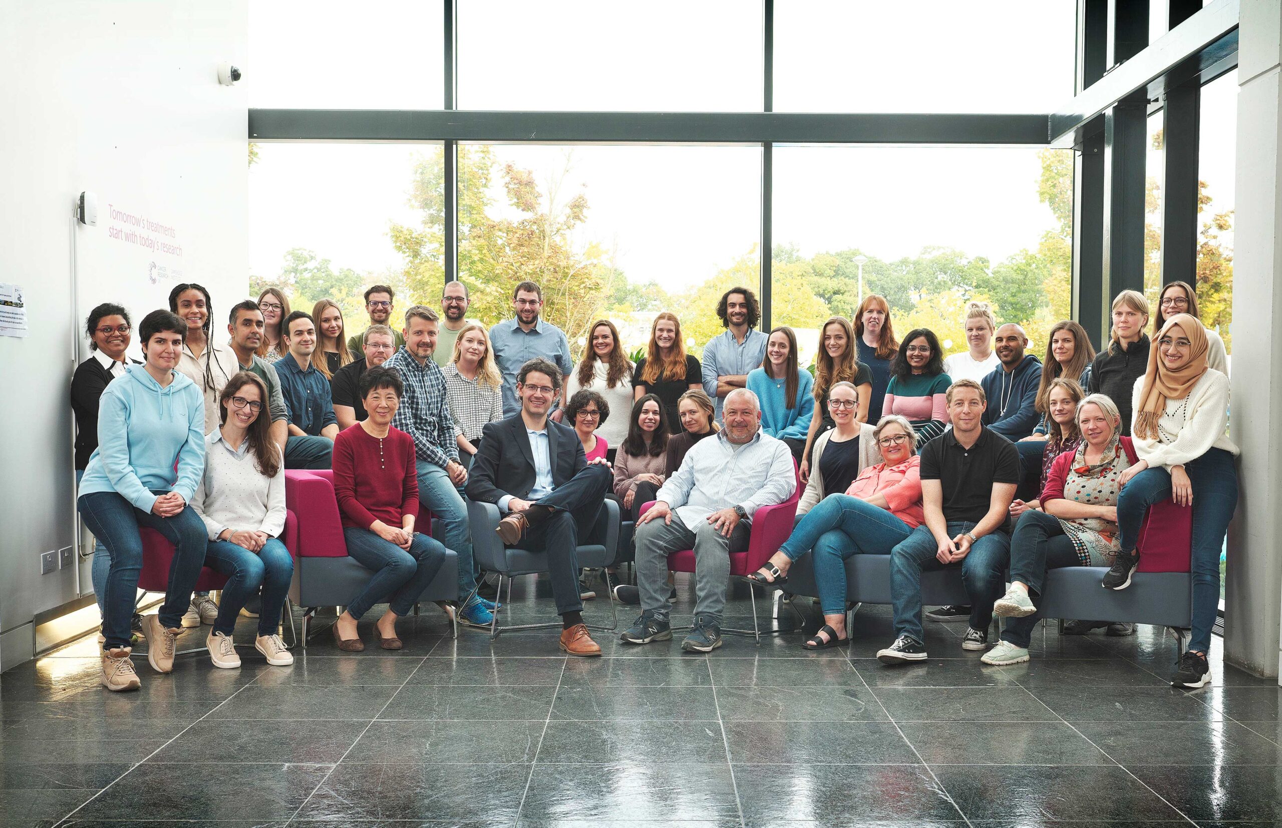
We are global team of experts from fields as diverse as medicine and astronomy, programming and molecular biology, and virtual reality (VR) and statistics. By combining existing techniques with entirely new approaches, IMAXT aims to build the first computerised 3D tumour that can be viewed in VR.
IMAXT: Imaging and Molecular Annotation of Xenografts and Tumours
To fully understand cancer, scientists need to know everything about a tumour – what types of cells are in it, how many there are, what they are doing and where they are located in the tumour. Having such a detailed picture of a tumour would allow scientists and doctors to develop new ways to diagnose and treat the disease, and new ways to stop it spreading and coming back.
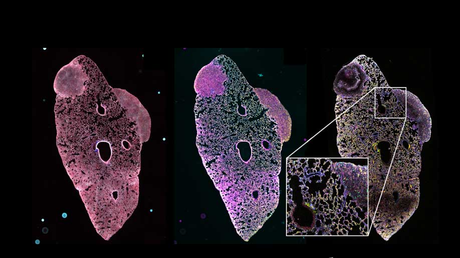
But getting such an accurate, precise picture of tumours is extremely difficult to do. So difficult that it’s not been done before. In this Cancer Research UK Grand Challenge project, scientists aim to build a 3D tumour that can be studied using virtual reality and which shows every single different type of cell in the tumour.
The project is on such a vast scale that a large, multi-national team has been assembled. It includes scientists from as far afield as the west coast of Canada, Ireland, Switzerland, UK and US. Each team member is an expert in their field, from astronomy to DNA sequencing, virtual reality to molecular biology, and statistics to medicine. This varied expertise will be crucial in allowing the team to combine established techniques, including DNA sequencing and imaging, with new technology they will invent and develop. The team will be led by Professor Greg Hannon who has likened the project to a mission to put a man on Mars.
The scientists aim to gather thousands of bits of information about every single cell in a tumour and use it to construct a 3D version that can be studied using virtual reality. This will allow everybody from scientists to patients to students to understand cancer in a new way.
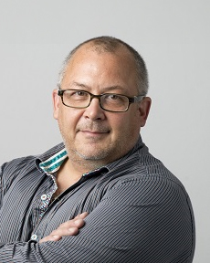
Professor Greg Hannon
Senior Group Leader