Watching the death throes of tumours
A clinical trial due to begin later this year will see scientists observing close up, in real time – and in patients – how tumours respond to new drugs.
There was a time when diagnosing and treating cancer seemed straightforward. Cancer of the breast was breast cancer, for example, and doctors could only choose treatments from a limited arsenal.
Now, the picture is much more complicated. A study published in 2012, led by Carlos Caldas, showed that breast cancer was actually at least ten different diseases. In fact, genome sequencing shows that even one ‘type’ of breast cancer differs between individuals.
While these developments illustrate the complexity of cancer biology, they also offer the promise of drugs tailored to an individual. Chemotherapy is a powerful, but blunt, instrument – it attacks the tumour, but in doing so also attacks several of the body’s other functions, which is why it makes patients so ill. The new generation of cancer drugs aim to make the tumour – and not the patient – sick.
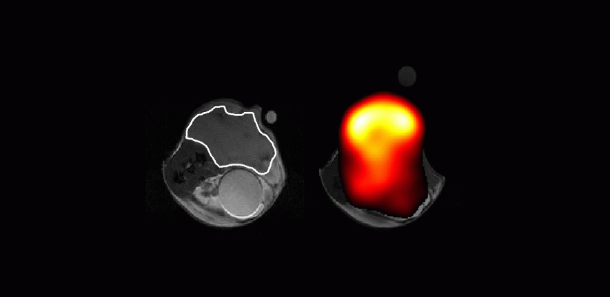
But telling if a patient is sick is easy; telling if the tumour is sick is more challenging. “Conventionally, one assesses whether a tumour is responding to treatment by looking for evidence of shrinkage,” explained Professor Kevin Brindle from the Cancer Research UK (CRUK) Cambridge Institute, “but that can take weeks or months. And monitoring tumour size doesn’t necessarily indicate whether it is responding well to treatment.”
Take brain tumours, for example. They can continue to grow even when a treatment is working. “The thing is that a tumour is not just tumour cells. There are lots of other cells in there, too.”
For some time now, oncologists have been interested in imaging aspects of tumour biology that can give a much earlier indication of the effect of treatment. Positron emission tomography (PET) can be used for this purpose. The patient is injected with a form (or analogue) of glucose labelled with a radioactive isotope. Tumours feed on the analogue and the isotope allows doctors to see where the tumour is.
An alternative technique that doesn’t expose the patient to ionising radiation is magnetic resonance imaging (MRI), which relies on the interaction of strong magnetic fields with a property of atomic nuclei known as ‘spin’. The proton spins in water molecules align in magnetic fields, like tiny bar magnets. By looking at how these spins differ in the presence of magnetic field gradients applied across the body, scientists are able to build up three-dimensional images of tissues.
In the 1970s, scientists realised that it was possible to use MR spectroscopy to see signals from metabolites such as glucose inside cells. “Tumours eat and breathe. If you make them sick, they don’t eat as much and the concentration of some cell metabolites can go down,” said Brindle.
Around the same time, scientists hit upon the idea of enriching metabolites with a naturally occurring isotope of carbon known as carbon-13 to help them measure how these metabolites are used by tissues. But carbon-13 nuclei are even less sensitive to detection by MRI than protons, so the signals are boosted using a machine developed by GE Healthcare, called a hyperpolariser, which lines up a large proportion of the carbon-13 spins before injection into the patient.
In 2006, Cambridge was one of the first places to show that this approach could be used to monitor whether a cancer therapy was effective or not. Combined with the latest genome sequencing techniques, this could become a powerful way of implementing personalised medicine. What’s more, because no radioactive isotopes are involved, an individual could be scanned safely multiple times.
“Because of the underlying genetics of the tumour, not all patients respond in the same way, but if you sequence the DNA in the tumour, you can select drugs that might work for that individual. Using hyperpolarisation and MRI, we can potentially tell whether the drug is working within a few hours of starting treatment. If it’s working you continue, if not you change the treatment.”
The challenge has been how to deliver the carbon-13 to the patient. The metabolite has to be cooled down to almost absolute zero (–273°C), polarised, warmed up rapidly, passed into the MRI room and injected into the patient. And as the polarisation of the carbon-13 nuclei has a half-life of only 30–40 seconds, this has to be done very quickly.
This problem has largely been solved and, with funding from the Wellcome Trust and CRUK, Brindle and colleagues will this year begin trialling the technique with cancer patients at Addenbrooke’s Hospital. If successful, it could revolutionise both the evaluation of new drugs and ultimately – and most importantly – the treatment of patients.
“Some people have been sceptical about whether we could ever get a strong enough signal. I’m sure we will. But will we be able to do something that is clinically meaningful, that is going to change clinical practice? That’s the big question we hope to answer in the coming years.”
Related News
See all news-
In Memoriam: Professor Greg Hannon (1964–2026)
9th April 2026
It is with profound sadness that we share the news of the passing of Professor Greg Hannon, who led the Cancer Research UK Cambridge Institute for over eight years.
Find out more -
Most detailed map of breast tissue changes reveals role of menopause in cancer susceptibility
31st March 2026
Scientists have created the most detailed map to date, comprising over 3 million cells, showing how breast tissue changes as women age – including dramatic changes during menopause.
Find out more -
Hannon Group joins global team to decode cancer’s dark proteome
4th March 2026
Prof Greg Hannon and his group joins global Cancer Grand Challenges team taking on the dark proteome challenge.
Find out more