New imaging technique spots prostate tumours starved of oxygen

A new imaging technique uncovers oxygen levels in prostate tumours and could lead to a non-invasive way to determine which tumours are more difficult to treat, according to a Cancer Research UK-funded study published in Theranostics.
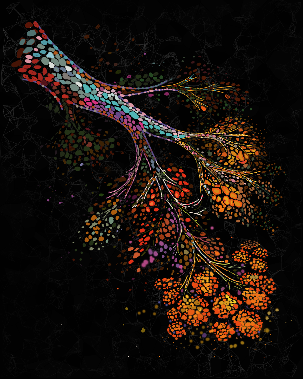
Scientists at the University of Cambridge created a new technique with an imaging device using a combination of light and sound to check the oxygen levels in prostate tumours in mice.
They gave mice a short burst of pure oxygen to breathe, and monitored how quickly and efficiently the extra oxygen reached different tumour areas through the blood supply.
Using the imaging device, the researchers were able to image the stronger response to oxygen by tumours with better blood vessels, which could give vital information on the quality of vessels.
This could help doctors find patients with harder to treat prostate cancers. Cancer cells supplied by poor quality blood vessels and low oxygen levels – called hypoxic conditions – are more resistant to drugs and radiotherapy.
Not only are cells in hypoxic conditions hardier and better at adapting to harsh conditions, making them more aggressive, but poor blood vessels also reduce the number of treatments like chemotherapy that can penetrate into the heart of the tumour.
Dr Sarah Bohndiek, scientist at the Cancer Research UK Cambridge Institute, said: “Our new imaging technique gives us a clearer picture of the heart of prostate cancer than we have ever had before.
“The tortuous nature of blood vessels can leave tumours starved of oxygen – making the cancer resistant to radiotherapy and chemotherapy and very difficult to treat. If we can translate this technology to the clinic, we could provide a non-invasive way to stratify men for treatment and monitor the effect of different therapies.”
Every year, about 46,500 men are diagnosed with prostate cancer and around 11,000 men die from the disease.
Professor Karen Vousden, Cancer Research UK’s chief scientist, said: “This new technique complements existing MRI and PET scans by helping us learn more about blood vessels within the tumour, which we haven’t been able to look at before in this way.
“It’s important that we develop better techniques to understand a patient’s individual tumour. This new tool gives insight into the tumour environment and could help separate the less aggressive forms of prostate cancer from the more difficult to treat.”
This research was made possible through the support of the Biological Resource Unit, Histopathology, Light Microscopy, and Imaging Core Facilities at the Institute.
Related News
See all news-

In Memoriam: Professor Greg Hannon (1964–2026)
9th April 2026
It is with profound sadness that we share the news of the passing of Professor Greg Hannon, who led the Cancer Research UK Cambridge Institute for over eight years.
Find out more -
Most detailed map of breast tissue changes reveals role of menopause in cancer susceptibility
31st March 2026
Scientists have created the most detailed map to date, comprising over 3 million cells, showing how breast tissue changes as women age – including dramatic changes during menopause.
Find out more -

Hannon Group joins global team to decode cancer’s dark proteome
4th March 2026
Prof Greg Hannon and his group joins global Cancer Grand Challenges team taking on the dark proteome challenge.
Find out more