First cancer patient in Europe scanned using new technique
The first cancer patient in Europe has been scanned with a revolutionary imaging technique that could enable doctors to see whether a drug is working within a day or two of starting treatment.
The patient is the first to take part in a new metabolic imaging trial of patients across a wide range of cancer types to be carried out by Cancer Research UK-funded scientists at Addenbrooke’s Hospital, part of Cambridge University Hospitals.
The study, which is funded by a Wellcome Trust Strategic Award, could show whether patients can stop taking drugs that aren’t working for them, try different ones and receive the best treatment for their cancer as quickly as possible.
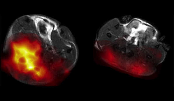
The rapid scan will allow doctors to map out molecular changes in patients, opening up potential new ways to detect cancer and monitor the effects of treatment.
The technique uses a breakdown product of glucose called pyruvate. The pyruvate is labelled with a non-radioactive form of carbon, called carbon 13 (C-13) which makes it 10,000 times more likely to be detected in a magnetic resonance imaging (MRI) scan. Pyruvate is injected into the patient and tracked as the molecule moves around the body and enters cells. The scan monitors how quickly cancer cells break pyruvate down – a measure of how active the cells are that tells doctors whether or not a drug has been effective at killing them.
Professor Kevin Brindle, co-lead based at the Cancer Research UK Cambridge Institute, said: “We’re very excited to be the first group outside North America, and the third group world-wide, to test this with patients and we hope that it will soon help improve treatment by putting to an end patients being given treatments that aren’t working for them. Each person’s cancer is different and this technique could help us tailor a patient’s treatment more quickly than before.”
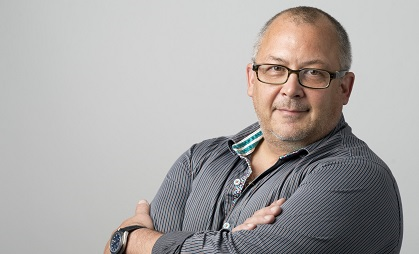
Dr Ferdia Gallagher, co-lead also funded by Cancer Research UK and based at the Department of Radiology at the University of Cambridge, said: “It’s fantastic that we can now try this technique in patients. We hope this will progress the way cancer treatment is given and make therapy more effective for patients in the future. This new technique could potentially mean that doctors will find out much more quickly if a treatment is working for their patient instead of waiting to see if a tumour shrinks.”
Dr Emma Smith, Cancer Research UK’s science information manager, said: “Finding out early on whether cancer is responding to therapy could save patients months of treatment that isn’t working for them. The next steps for this study will be collecting and analysing the results to find out if this imaging technology provides an accurate early snapshot of how well drugs destroy tumours.”
Related News
See all news-
In Memoriam: Professor Greg Hannon (1964–2026)
9th April 2026
It is with profound sadness that we share the news of the passing of Professor Greg Hannon, who led the Cancer Research UK Cambridge Institute for over eight years.
Find out more -
Most detailed map of breast tissue changes reveals role of menopause in cancer susceptibility
31st March 2026
Scientists have created the most detailed map to date, comprising over 3 million cells, showing how breast tissue changes as women age – including dramatic changes during menopause.
Find out more -
Hannon Group joins global team to decode cancer’s dark proteome
4th March 2026
Prof Greg Hannon and his group joins global Cancer Grand Challenges team taking on the dark proteome challenge.
Find out more