In Situ Hybridisation and Spatial Omics Symposium
20–21 May 2026
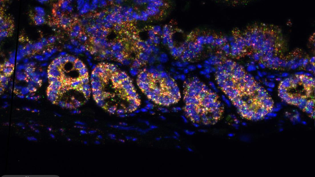
Join us at the Cancer Research UK Cambridge Institute for a two-day symposium exploring the latest advances in in situ hybridisation and spatial omics – technologies that are transforming our understanding of cellular architecture, gene expression, and tissue microenvironments.
Sessions:
- Advancing In Situ Hybridisation Technologies
- ISH in Disease Diagnostics and Therapeutics
- 3D Models and ISH in Complex Biological Systems
- The Future of Spatial Biology: Integrating ISH with Multiomics and AI
Event Details
Location: Cancer Research UK Cambridge Institute
Dates: 20-21 May 2026
Format: In person with keynote talks, panel discussions, poster sessions and networking opportunities. Formal symposium dinner at Homerton College, Cambridge on the 20th May.
Registration
Registration is now open.
Early Bird Rate:
Symposium only £200
Symposium with dinner £255
After 13 April 2026:
Symposium only £220
Symposium with dinner £275
Confirmed speakers
Professor Ilan Davis – University of Glasgow
Ilan has been at the forefront of demonstrating that pattern formation, neural stem cell development and synaptic plasticity, involve crucial regulation by post-transcriptional regulation. His lab discovered the first example of Dynein transporting mRNA along microtubules during pattern formation in Drosophila embryo and oocyte and demonstrated that mRNA is statically anchored by the motor. They also characterised how phase transition processing bodies triage the fate of mRNA for immediate translation in oocytes or storage for later translation in the embryo. Current research interests include Drosophila neuroscience, viruses and RNA, and microscope technology development.
Professor Long Cai – Caltech, US
My lab focuses on developing methods and tools for the field of spatial genomics. We showed that a large number of mRNAs can be profiled in single cells in their native environments in tissues. We accomplished this by developing a sequential barcoding method with sequential fluorescence in situ hybridization (seqFISH) (Lubeck et al, Nat Meth 2014). The multiplexing capacity of this method scales exponentially, allowing the entire transcriptome of over 10,000 genes to be barcoded (Eng et al, 2017 and Shah et al, Cell 2018, Eng et al, Nature 2019) . Overcoming the bottleneck of coding for different mRNAs in cells opens the door for answering a large range of problem in spatially heterogeneous tissues that are inaccessible with existing methods. We have been translating this method to solve fundamental questions in developmental biology and neuroscience.
Building upon seqFISH, we developed a method called Memory through Engineered Mutagenesis with Optical In situ Readout (MEMOIR). This method allows us to record cellular events into the genome of cells using CRISPR based genome editing tools, and readout of this information in situ using seqFISH. We have shown experimentally that MEMOIR can track lineage trees with single cell information.
Recently, we demonstrated synaptic MEMOIR, which enables mapping of synaptic connections in organisms. Synaptic MEMOIR has the potential to integrate connectomics with spatial genomics and provide a powerful tool in understanding cell identity and fate decisions are fundamental to a wide range of questions in neuroscience, developmental biology and human diseases.
Dr Ryan MacDonald – University College London
The overarching research aim of my laboratory is to understand how a healthy eye is built and maintained throughout life. More specifically, we are interested in how glial cells, the major support cells in the nervous system, are patterned and shaped during development to support neurons. We are also interested in what happens when the intricate glia-neuronal relationship breaks down due to increasing age or disease.
The retina is the light sensitive part of the eye that allows you to see. We use the zebrafish retina as a model as it contains the same neuron types and glial cells as the human eye. The zebrafish embryo is an incredible system to study development – it is transparent, we can label each cell with specific fluorescent markers and use time-lapse confocal microscopy to watch eye development happen in real time in a living fish! In addition to imaging we use CRISPR/Cas9 mutagenesis, RNA-sequencing and molecular biology to uncover and explore fundamental mechanisms of glial biology.
We are now interested in determining the cellular and molecular mechanisms regulating glial morphogenesis, with a particular focus on the consequences of disrupted glial contacts on neuronal function.
Dr Brian Beliveau – University of Washington
The Beliveau Lab is focused on building robust and scalable enabling technologies to study the organization of chromosomes in 3D space, the interactions they participate in at the inter- and intra-chromosomal level, and the associated RNAs and proteins that occupy functionally relevant sites. The motivation for this work is to better understand the mechanisms by which the organization and composition of genomic intervals relevant for health and disease impact the essential DNA transactions of transcription, replication, and repair. We also are committed to building ecosystems supported by open-source software, low-cost hardware, and extensive documentation to democratize the adoption of advanced single cell and spatial approaches in order to facilitate their application in a broad range of research settings.
Professor Hernan Garcia – University of California, Berkeley
Hernan G. Garcia is an Associate Professor in the Departments of Molecular & Cell Biology and Physics at UC Berkeley, where his research seeks a predictive, quantitative understanding of how cells make fate decisions during embryonic development. His group combines live imaging of transcription in intact embryos with theoretical modeling to uncover how regulatory DNA sequences encode precise spatiotemporal patterns of gene expression. Using the fruit fly as a primary model system, they develop physics-inspired models that generate testable predictions and design experiments to directly confront them. A major current effort focuses on scaling in situ and imaging-based genomic dissections to unprecedented throughput, enabling the analysis of tens of thousands of embryos in single experiments. Together, these approaches aim to transform gene regulation from a descriptive to a predictive science, with implications for developmental biology, genomics, and non-equilibrium physics.
Dr Boye Schnack Nielsen – Bioneer, DK
The team at Bioneer is currently exploring the use of 3D models for drug testing. In addition to growing co-cultures of cancer cell lines, fibroblasts and immune cells, we work with organoid development from iPSCs. Basic characterization of 3D models is key to reach their translational application. Drug testing include standard cytotoxic assays, involving chemotherapeutic agents and immune-oncology applications.
Dr Dario Bressan – University of Cambridge
I focus on technology development in the field of spatial omics, applied to cancer research. In particular our laboratory focuses on the integration of multiple technologies to produce multi-omic, deep datasets that can assist with the investigation of tumour heterogeneity and tumour-microenvironment interactions. I direct the Spatial Profiling and Annotation Centre of Excellence, a collaborative laboratory sponsored by the Cancer grand Challenges initiative providing access to cutting-edge spatial omics technologies to research projects from all over the world
Professor John Le Quesne – CRUK Scotland Institute
The Le Quesne group integrate high-parameter spatial biology with clinical pathology to decode the landscape of solid tumours, focussing on respiratory system malignancies. Our research is centred on two converging pillars: advanced technology development and multimodal AI interpretation.
We specialize in the optimization of highly multiplexed protein and RNA detection, utilizing Akoya, Lunaphore, and multiplexed RNAscope (ISH), alongside spatial transcriptomics such as 10x Xenium. By quantifying axes of translational control and cellular stress in situ, we explore how single-cell reprogramming drives aggressive tumour phenotypes.
The development of self-supervised AI frameworks that joint-model morphology and molecular phenotype from these data-rich ISH and proteomic datasets is central to our work. These approaches allow us to discover novel “lethal” morphologies and prognostic spatial patterns that escape traditional expert interpretation and classical digital image analysis.
Dr Madeline Lancaster – MRC Laboratory of Molecular Biology
Research in the Lancaster lab focuses on human brain development using stem cells to generate brain organoids that allow modelling of human brain development in vitro. The laboratory studies the most fundamental differences between human brain development and that of other mammalian species. The lab also studies cellular mechanisms underlying neurodevelopmental disorders such as autism and intellectual disability.
Professor Thomas Walter – Mines Paris / Institut Curie
Thomas Walter’s research focuses on developing methods in artificial intelligence and computer vision for the life sciences. He has pioneered approaches to computational phenotyping for high-content screening, characterizing phenotypic responses to perturbation experiments through the analysis of cellular and nuclear morphology. His work has also addressed the analysis of RNA localization patterns. A second major application area is computational pathology, where he develops predictive models for whole-slide images to infer clinically relevant variables such as specific mutations, transcriptomic subtypes, prognosis, or treatment response. More recently, his research has expanded to spatial transcriptomics, with contributions to cell segmentation in image-based spatial transcriptomics and to cross-modality prediction methods that link morphological phenotypes with transcriptomic and proteomic measurements.
Dr Raza Ali – Cancer Research UK Cambridge Institute
Our aim is to develop a deeper understanding of heterocellular tumour ecosystems and to translate our discoveries for patient benefit.
Tumours are mixtures of different cells. The phenotypes of these cells and their interactions direct spatial arrangements and underpin disease progression. We characterise cells in their native context within intact tissues by precisely defining their phenotype and spatial context at multiple scales. Taking a ‘systems’ view of tumours as communities of diverse interacting cells will provide insights into cancer biology, and lead to new ways of diagnosing and treating cancer.
To generate these detailed snapshots of human tumour ecosystems we use multidimensional molecular tissue imaging (imaging mass cytometry) to simultaneously map between forty and fifty molecules with subcellular spatial resolution. We use computational tools to extract cellular phenotypic data and represent cell-cell interactions as spatial networks. In landmark studies we were first to link these multi-dimensional tumour phenotypes to somatic genomic alterations (Danenberg et al Nature Genetics 2022; Ali HR et al Nature Cancer 2020). We also investigate how these new insights can be translated to conventional digital pathology.
Our focus is breast cancer, still a major cause of premature death. New, effective diagnostic tools and treatments are urgently needed. We aim to identify the spatial determinants of disease progression, treatment response, and resistance (Wang et al Nature 2023).
Dr Richard Tyser – Cambridge Stem Cell Institute
Our team explores how the mammalian heart begins to form and function during embryonic development. The heart is the first organ to form and function during embryogenesis, essential in providing the developing embryo with sufficient oxygen and nutrients.
We use a combination of imaging and molecular based approaches to characterise cardiac progenitor cell populations, in both the human and mouse. Using this insight, we examine the mechanisms which regulate how cardiac progenitors differentiate to give rise to the functional beating heart.
We are particularly focused on understanding how the onset of contraction influences heart morphology and cardiomyocyte differentiation. As well as addressing questions of fundamental biological significance, our research aims to augment therapeutic approaches to treat disease: by establishing the underlying causes of disease as well as providing a blueprint for regenerative strategies on how best to treat them.
Prof Udeni Balasuriya – School of Veterinary Medicine, Louisiana State University
My research program at LSU combines classical and contemporary laboratory methods, including advanced spatial biology techniques, to enhance diagnostics and deepen our understanding of virus–host interactions involving veterinary and zoonotic viral pathogens.
We develop and implement highly sensitive real-time and digital PCR assays, as well as tissue-based methods such as immunohistochemistry (IHC) and in situ hybridization (RNAscope™), to detect and localize viral nucleic acids and proteins (viral and cellular markers) in host tissues.
A key focus of our work is to define cellular tropism and viral pathogenesis, including persistence, and to elucidate host barriers to cross-species infection through spatially resolved analyses. Furthermore, these contemporary techniques will be combined to study the mechanisms underlying retroviral persistence, enabling a deeper understanding of how they evade the host immune system.
By integrating molecular diagnostics with spatial omics-inspired methodologies, my research contributes to One Health initiatives at the animal–human interface.
Prof Rong Fan – Department of Engineering, Yale University
Dr. Rong Fan’s current research at Yale centers on developing and applying cutting-edge single-cell and spatial multi-omics technologies to transform our understanding of human tissues in health and disease.
His lab creates highly multiplexed platforms that quantify proteins, gene expression, and epigenetic states at single-cell resolution within intact tissue contexts.
Key innovations include high-throughput single-cell secretome profiling, genome-scale spatial transcriptomics and proteomics, and the first spatial epigenomics methods such as spatial-CUT&Tag and spatial-ATAC-seq, as well as integrated spatial epigenome–transcriptome co-sequencing. These tools reveal tissue architecture, cellular interactions, and immune microenvironments with unprecedented molecular detail.
By enabling quantitative, spatially resolved molecular analysis directly in clinical specimens, Dr. Fan’s work advances digital pathology, biomarker discovery, and mechanistic insights into development, cancer, and immune-mediated diseases.
Our Sponsors
We would like to thank all of our sponsors, who help to make this event possible!


Organising Committee
Professor Scott Fraser – President, Dynamic Imaging, Biohub-SF, US
Scott Fraser has a long-standing commitment to quantitative biology, applying the tools of chemistry, engineering, and physics to problems in biology and medicine. His personal research centers on imaging analyses of intact biological systems, with an emphasis on early development, organogenesis, and medical diagnostics. The Biohub-SF which he leads is combining frontier AI with frontier biology/technology to understand cell and protein dynamics, using multimodal and multiscale imaging at scale.
Dr Julia Jones – Scientific Manager (ISH), Histopathology/ISH Core Facility, CRUK Cambridge Institute
I am responsible for the running of the In Situ Hybridisation (ISH) service within the histopathology core facility at CRUK-CI, with a focus on providing high-quality ISH services to support scientific research. During the 19 years since the CI opened, the ISH service has evolved from manual mRNA detection using radiolabeled riboprobes to the latest generation of manual and automated RNAscope assays from ACD for the detection of a multitude of RNA species. In addition, we offer a DNA FISH service. We work closely with CI scientists and external collaborators to design, run, and analyze all manner of in situ hybridisation projects. For our customers that want to gain a deeper understanding of gene expression and protein localization in their samples, we offer a multi-modal detection service for RNA and protein analysis on the same tissue section. Our goal is to deliver reliable results to help our scientists achieve their research goals and advance their scientific discoveries.
Dr Boye Schnack Nielsen – Principal Scientist, Bioneer, DK
The team at Bioneer is currently exploring the use of 3D models for drug testing. In addition to growing co-cultures of cancer cell lines, fibroblasts and immune cells, we work with organoid development from iPSCs. Basic characterization of 3D models is key to reach their translational application. Drug testing include standard cytotoxic assays, involving chemotherapeutic agents and immune-oncology applications.
Dr Dario Bressan – Head, Spatial Profiling and Annotation Centre of Excellence (SPACE), CRUK Cambridge Institute
I oversee a centre of excellence dedicated to the development, optimization, integration and application of spatial “omics” technologies to cancer research
Dr Aldo Ciau Uitz – Senior Scientist, Biomodal
Dr Will Howat – Vice President of Validation & Technical Quality, Abcam