Pre-clinical imaging
Applying imaging and spectroscopic methods to anatomy, function, and metabolism.
We provide a range of state-of-the-art pre-clinical imaging modalities and high-resolution NMR spectroscopy, maintaining close links with the Department of Radiology’s clinical research. Modalities include bioluminescence, fluorescence, ultrasound, MRI, PET, SPECT and CT, as well as photoacoustic imaging, whose development is the focus of one of the CI’s research groups. We additionally support the SARRP image-guided X-ray irradiators. Data acquired for external collaborators can be provided via an XNAT image database.

Dr Dominick McIntyre
Equipment
Bioluminescence and fluorescence in-vivo imaging systems
IVIS Spectrum and IVIS Lumina LT imaging systems (Perkin Elmer) are available for whole-animal in vivo photonic imaging. These can perform sensitive and relatively high-throughput in vivo bioluminescence imaging (BLI), in conjunction with luciferase labelled cells or tissues. Typical scans take less than one minute and up to five subjects can be imaged at a time. Both systems can also perform fluorescence imaging, including spectral unmixing for simultaneous imaging of multiple fluorescent reporters.
Additionally, we have a Licor Pearl Trilogy that can image only one subject at a time for a restricted range of fluorescent reporters, but has extremely low fluorescent background, giving higher sensitivity at the expense of lower throughput.
Optoacoustic imaging
We have two iThera optoacoustic imaging systems that enable access to the high contrast of optical imaging with the spatial resolution and penetration depth of ultrasound.
The MultiSpectral Optoacoustic Tomography (MSOT) system is capable of imaging between 680 and 950 nm, at a repetition rate of 10 wavelengths per second, enabling dynamic monitoring of oxygenation and contrast agent uptake. The Raster Scanning Optical Mesoscopy (RSOM) provides much higher spatial resolution of 20µm, using 532nm excitation and limited to a depth of a few millimetres.
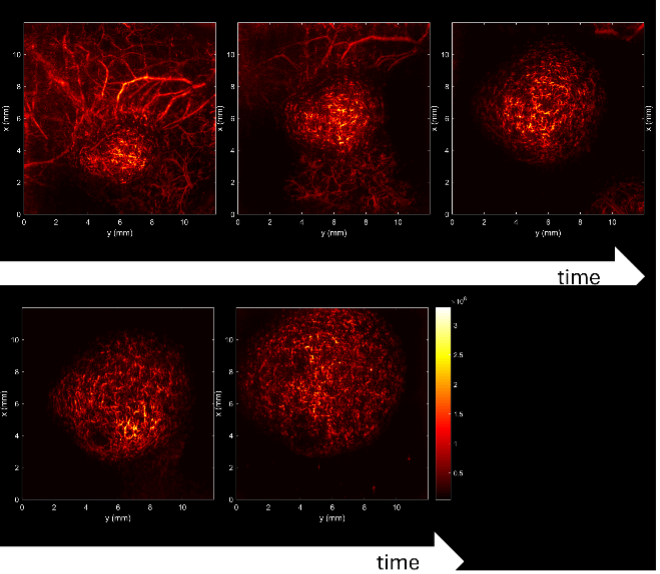
The image shows a series of blood vessel images from the RSOM, captured from the same model tumour at 2-3 day intervals as it grows and develops more complex vasculature (courtesy Dr M.-E. Oraiopoulou)
Magnetic resonance imaging
We have two MRI systems with integrated animal monitoring, cardiac and respiratory gating, heating, and isoflurane anaesthesia.
The 9.4T system has a modern Bruker Neo console and higher sensitivity from its higher magnetic field strength. The 7T, equipped with an older Agilent console, is more suitable for techniques such as echo-planar imaging due to the smaller susceptibility effects at this field.
Both perform 1H MRI and multi-nuclear MRS, including deuterium metabolic imaging. Improved 1H MRS methods that minimise chemical shift artefacts have been implemented. The core is developing quantitative MT-MRI and motion-insensitive DW-MRI methods for tumours in the abdomen, which are subject to respiratory and cardiac motion.
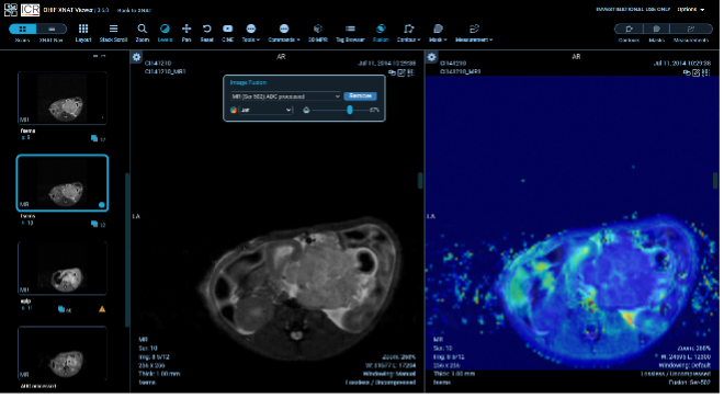
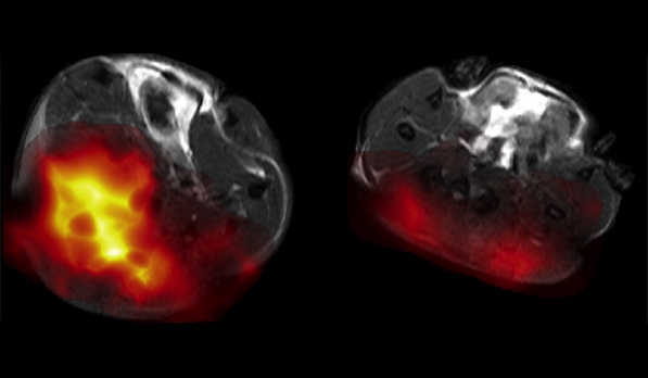
Additionally, we have an EchoMRI body composition system, providing a rapid readout of lean and fat mass, useful for conditions such as cachexia. Figure: T2-weighted FSE image of tumour and same image with apparent diffusion coefficient colourmap overlaid, showing necrosis not apparent in the T2W image
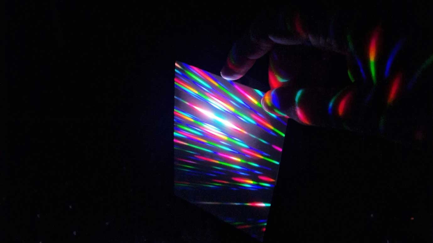
High-resolution NMR spectrometer
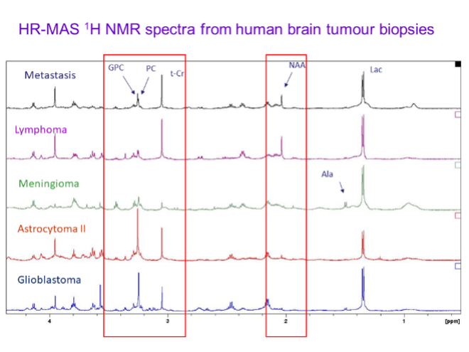
The Bruker 600 MHz Avance NMR spectrometer provides detailed information on the chemical content of samples. Cells, tissue biopsies (including human tissues with CL2 preparation), metabolites extracted from cells, and body fluids such as blood can be measured, using multinuclear probes including a high-resolution magic-angle (HR-MAS) spinning probe to obtain narrow spectral lines from biopsies. Metabolomics, structural studies using multidimensional methods, and analysis of uptake and metabolism of anti-cancer drugs are possible.
The figure shows HR-MAS spectra of brain tumours; red boxes denote the regions with largest differences between tumour types.
Hyperpolarized 13C MR imaging
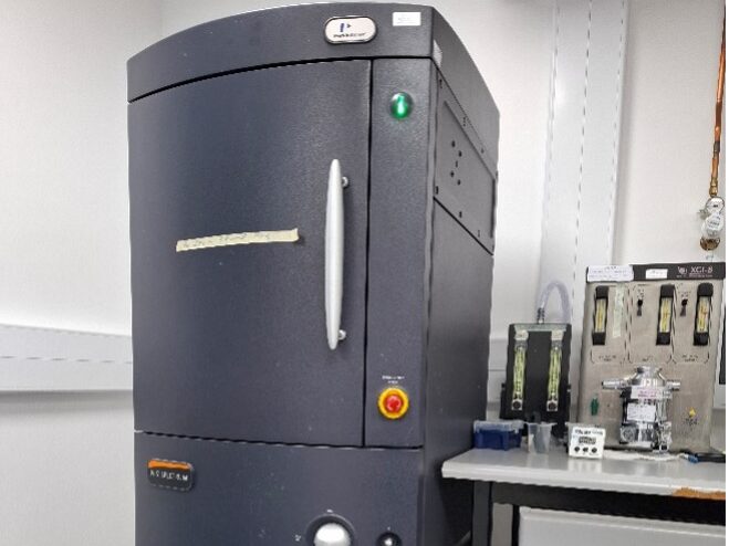
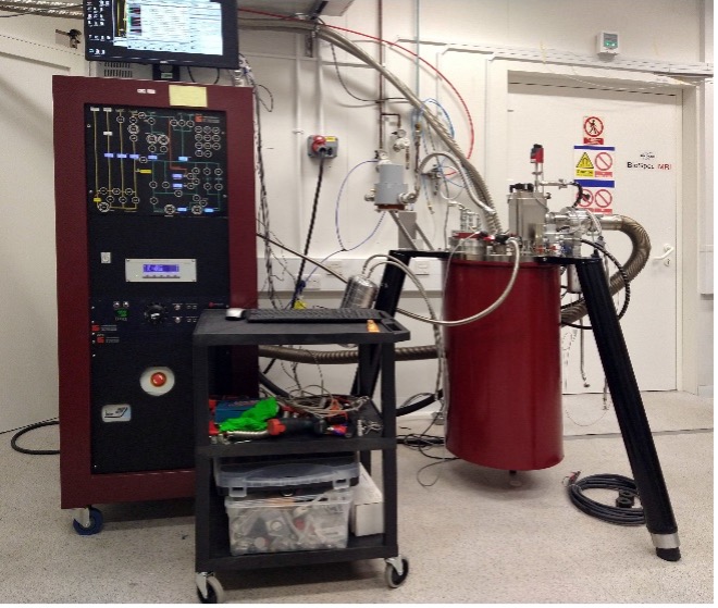
We are developing hyperpolarized 13C MR imaging as a novel cancer imaging tool. This allows imaging of the spatial distribution of injected 13C-labelled substrates and the metabolites formed from them. This has been used to image tumour response to chemotherapy, tumour pH, and necrosis. The facility has a cryogen-free DNP polariser (see image) capable of polarising multiple samples simultaneously, at a variable field of up to 7T.
Radionuclide imaging: PET and SPECT
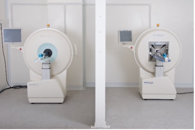
NanoScan PET/CT (Mediso, Hungary) and NanoSPECT (Bioscan, USA) are available for multimodality radionuclide imaging. These systems offer the greatest sensitivity of any in vivo imaging modality and can provide non-invasive assessment of pharmacological (target tissue exposure, target engagement and functional activity) and biological processes (blood flow, perfusion and metabolism). These scanners have nanomolar sensitivity and good resolution (~0.4 mm for SPECT and ~1 mm for PET) and so are ideal for small animal imaging.
Radiochemistry
The PET, CT, and SPECT imaging facility is equipped with in-house production capabilities for radioligands using a variety of radionuclides, including 99mTc, 111In, and 123I for Single Photon Emission Computed Tomography (SPECT), as well as 68Ga and 89Zr for Positron Emission Tomography (PET).
Our Core maintains a long-standing collaboration with other research institutions, such as the Molecular Imaging Chemical Laboratory at the University of Cambridge, and clinical production units like the Radiopharmacy and Radiopharmaceutical Production Unit at Addenbrooke’s Hospital. These partnerships enhance our radioligand production capabilities by incorporating additional radionuclides like 11C, 18F, and 68Ga for PET/CT.
The molecular probes currently being studied include [11C]acetate, [18F]FLT, [18F]FMISO, [18F]FET, [18F]C2Am, and [89Zr]-ligands, which are used for imaging processes such as fatty acid synthesis, proliferation, hypoxia, amino acid uptake, cell death, and CAR-T cell trafficking.
Ultrasound
Ultrasound provides rapid volumetric measurements of tumours deep in the body, providing the most efficient way to track tumour growth and regression with treatment for tumours that do not express bioluminescent or fluorescent proteins. It can also be used for image-guided injections.
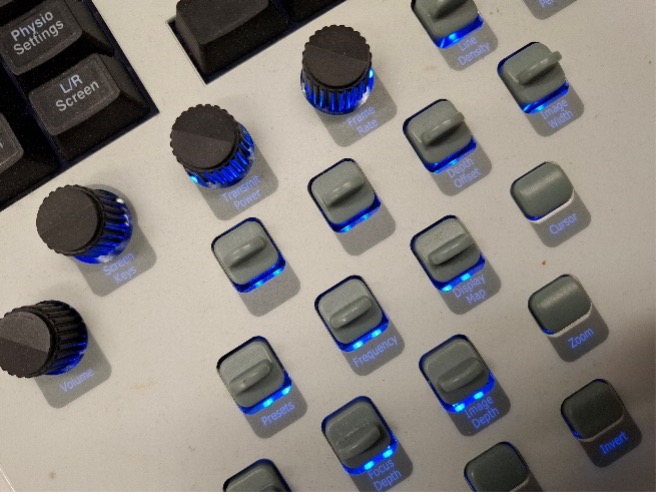
We have two Visualsonics systems, a modern Vevo F2 and an older Vevo 2100, both capable of 2D and 3D structural imaging and a range of Doppler-based flow measurements. Image: Vevo 2100 control panel
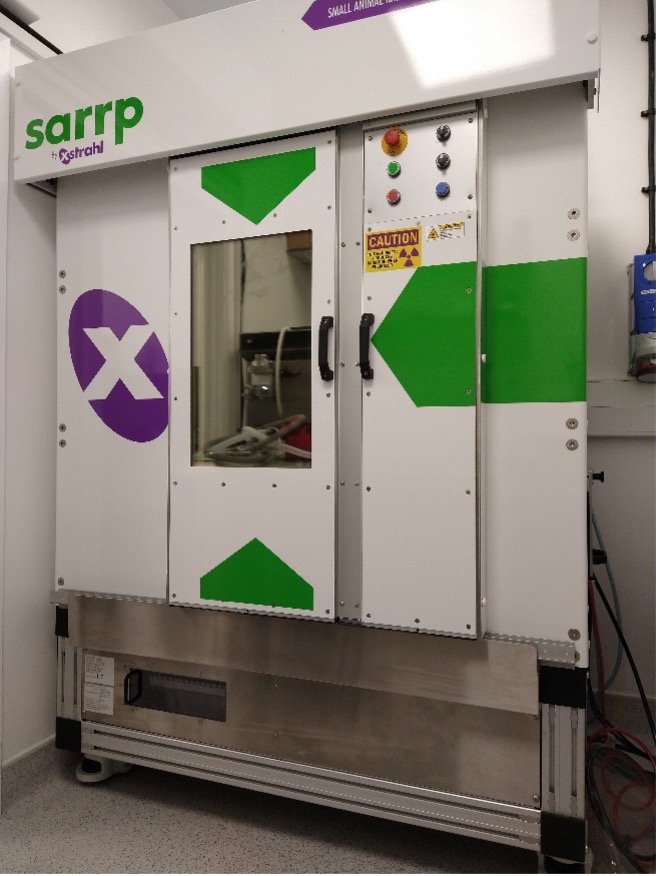
SARRP X-ray image-guided irradiator
The SARRP irradiator, funded by CRUK’s RadNet program, employs an X-ray source for both CT imaging and targeted radiotherapy. This allows precisely registered targeting of treatment, using beams as narrow as 0.5mm. Planning software and motorised gantry and bed enable high doses to be delivered to the tumour while sparing normal tissues.
The Imaging Core are engaged in a collaborative multicentre project to improve performance by merging the CT data with images from other modalities that better define the tumour limits.
Preclinical imaging textbooks
If you are planning to use our facilities, and would like a better understanding of how preclinical imaging works, we would suggest the following resources:
The Basics of Visualizing, Analyzing, and Reporting Preclinical PET/CT Imaging Data
From Signals to Image: A Basic Course on Medical Imaging for Engineers
Biomedical Signal and Image Processing

Related News
See all news-
Imaging magnetised molecules could predict drug resistance in breast cancer patients
30th September 2020
New research suggests that a scanning technique, called carbon-13 hyperpolarised imaging, could be used to predict treatment response in breast cancer patients.
Find out more -
New imaging technique spots prostate tumours starved of oxygen
24th August 2017
A new imaging technique uncovers oxygen levels in prostate tumours and could lead to a non-invasive way to determine which tumours are more difficult to treat.
Find out more -
First cancer patient in Europe scanned in Cambridge using new technique showing whether drugs work
11th April 2016
The first cancer patient in Europe has been scanned with a revolutionary imaging technique that could enable doctors to see whether a drug is working within a day or two of starting treatment.
Find out more
Laboratory Efficiency Assessment Framework (LEAF)
Imaging contributed to the Institute’s LEAF Silver accreditation, see the Sustainability webpage for more information.