Histopathology and In Situ Hybridisation
Facilitating all aspects of histopathology including processing, embedding, sectioning, histochemical staining, tissue microarray building, immunohistochemistry, in situ hybridisation, whole slide scanning, and image analysis.
The ‘Histocore’ provides routine histological processing, tissue sectioning, immunohistochemistry (IHC), in situ hybridisation (ISH), and whole slide scanning services to Institute researchers (and external users where capacity allows). Histological services include the capability to process tissue samples and cell lines into frozen, paraffin or vibratome formats for sectioning and staining. The Core constructs tissue microarrays (TMAs) for users and trains in the use of Laser Capture Microdissection (LCM). We also offer image analysis services, so can offer a complete end-to-end service if required.

Jo Arnold
Head of Histopathology
Services
Routine Histology
The routine histology lab provides paraffin processing and embedding of pre-fixed tissue samples and work with researchers to orientate tissues in the best way for their needs. To a more limited degree we also carry out freezing of tissues, although it is recommended that tissues are frozen at source for best preservation.
In terms of sectioning, we offer standard and special paraffin sectioning services on our suite of microtomes, frozen sectioning on our cryostats and vibratome sectioning also. We can train researchers to use the cryostats or vibratome themselves for sectioning if preferred.
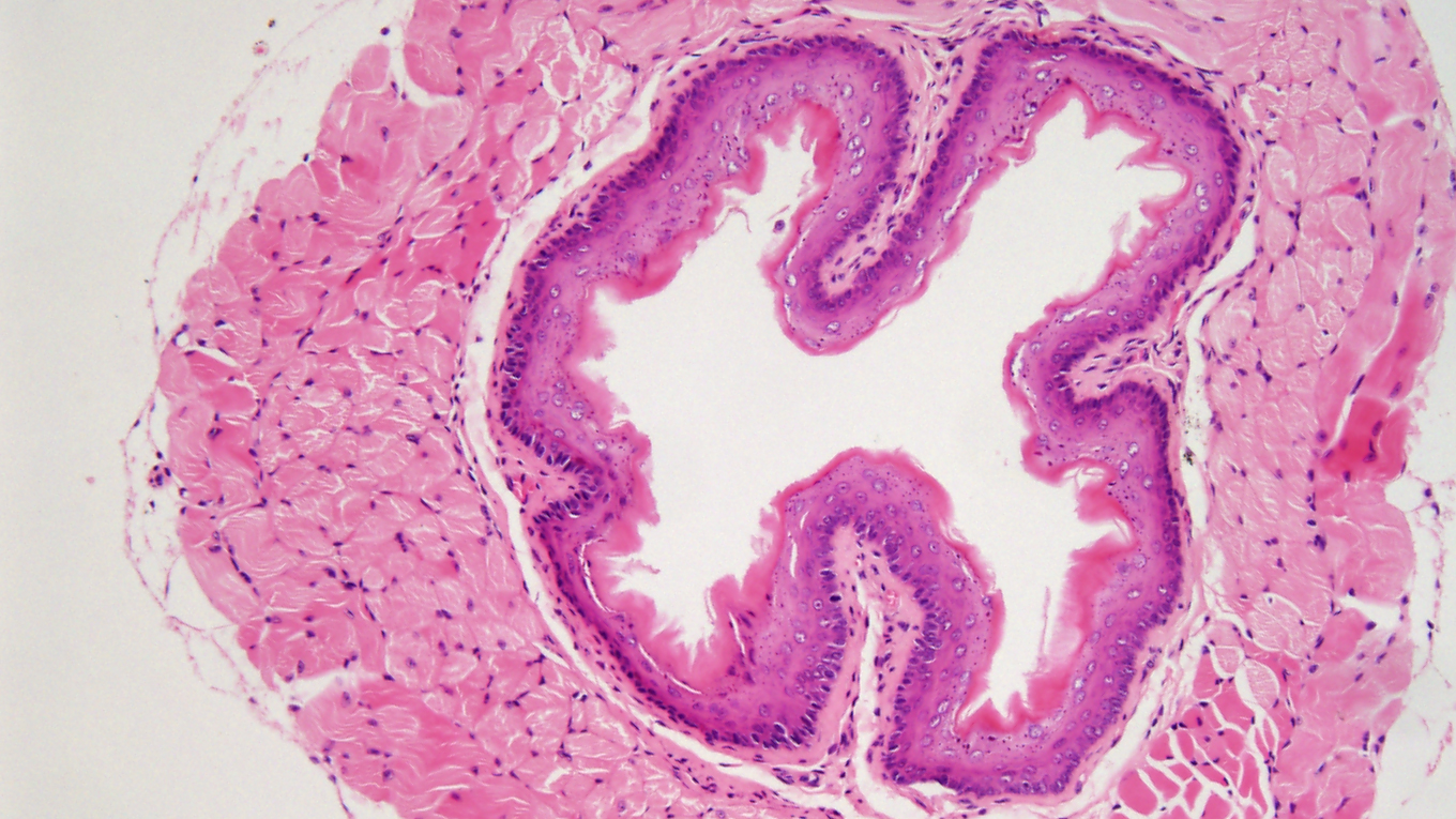
In addition to the standard haematoxylin and eosin (H&E) histochemical stain always recommended for QC and morphological assessment, the facility performs ‘special staining’, such as Masson’s Trichrome (to differentiate collagen and muscle fibres), Periodic-Acid-Schiff (to demonstrate glycogen), Alcian Blue (to visualise acidic epithelial and connective tissue mucins) and Congo Red (for the identification of amyloids). Tissue micro-array (TMA) building services are also available to users of our facility, starting with a project initiation meeting with our experts to discuss options.
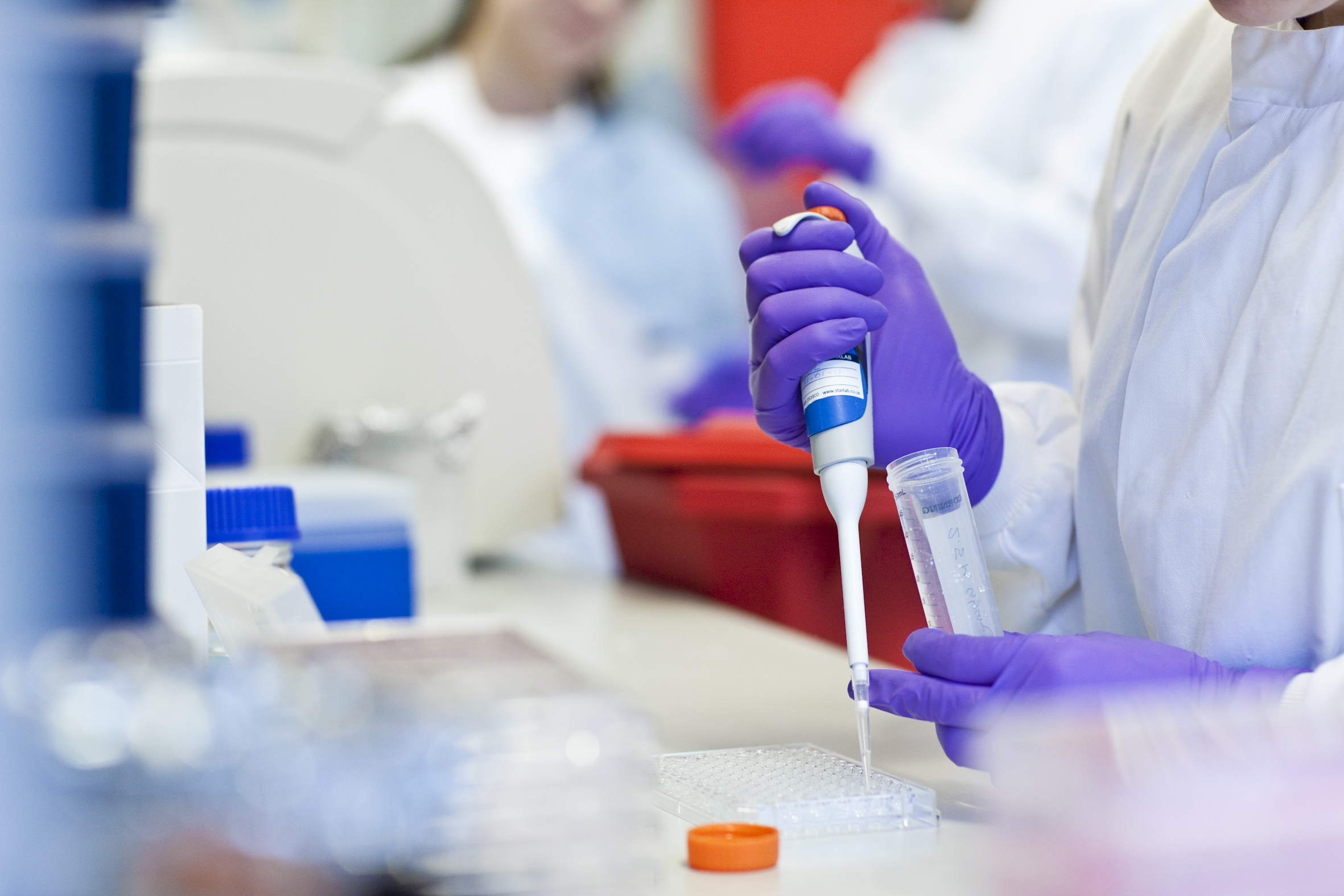
Immunohistochemistry
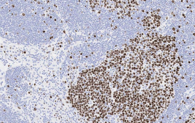
Immunohistochemistry (IHC) is the process of using antibodies specifically designed to bind to proteins of interest in tissue sections, which are then detected using enzyme mediated methods.
The facility utilises the Leica Bond systems for all IHC requests. This is a fully automated platform with an average turnaround time of 3 hours for a standard run. For human, murine, and rat tissue, we use an anti-mouse/anti-rabbit polymer-HRP detection system with DAB as the final substrate, and a haematoxylin counterstain.
We can detect other species of primary antibody by linking ancillary secondary antibodies into the polymer kit. We also carry out immunofluorescence (IF), dual IHC/special stains, TUNEL staining and a variety of multiplexing assays within this service area.
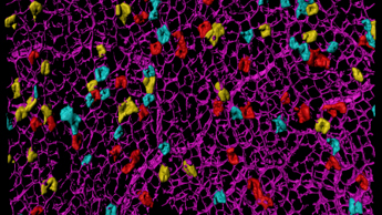
In Situ Hybridisation
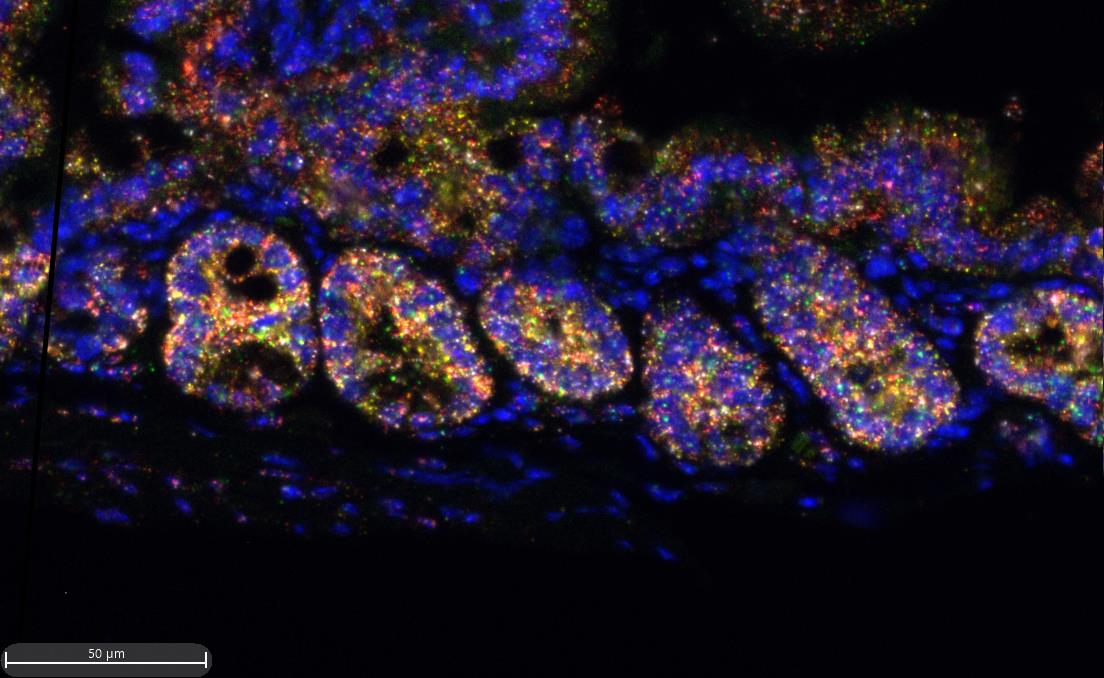
In situ hybridisation (ISH) is the process of detecting RNA or DNA species by means of hybridising a detectable complementary probe. The current method for detecting mRNA and non-coding RNA is using RNAscope technology from Advanced Cell Diagnostics (ACD). We can perform up to 4-plex ISH, fully automated on the Leica Bond Rx and up to 12-plex ISH using the manual HiPlex kit.
We also offer:
- BaseScope from ACD which can be utilised for detection of SNVs as well as exon junctions and low abundance targets.
- RNAscope Plus for the detection of 1 small RNA (microRNA, small interfering RNA, antisense oligonucleotides) plus up to 3 mRNA targets on the Leica Bond Rx.
- HiPlex assay from ACD for detection of 12 targets on one tissue section. This is performed manually in 3 rounds of 4 targets each. The slides are imaged on the Akoya Polaris between each round and then stripped of fluorophores before detection of the next 4 targets. Images are then ‘stacked’ and fused.
- DNA FISH using commercially sourced probes. We currently offer X/Y FISH, human/mouse pan centromeric FISH for xenograft models as well as other human DNA probes such as TP53.
- Integrated Codetection Workflow (ICW) from ACD to detect RNA and protein on the same slide using the Leica Bond Rx platform.
Scanning & Image Analysis
There are three whole slide scanners in the Core, run as a service only; the Leica Aperio AT2, the Zeiss Axioscan Z1, and the Akoya PhenoImager HT.
The Aperio is a fast scanner, 2 min/slide, dedicated to the image capture of H&E, special stained and IHC/ISH (chromogenic) stained slides, at 20x or 40x resolution.
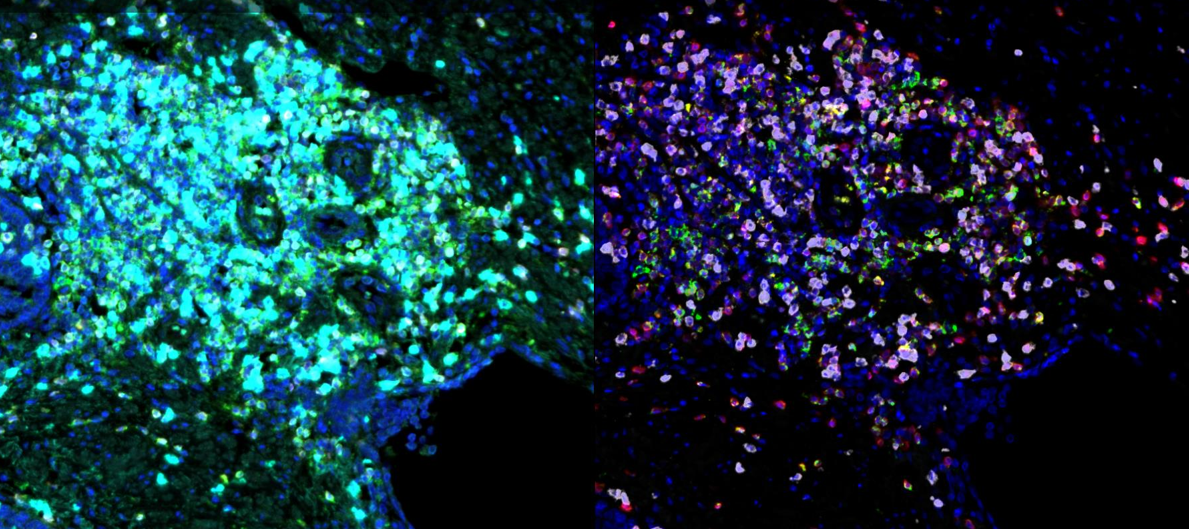
The Akoya PhenoImager HT is a brightfield and fluorescent multispectral scanner, capable of up to 40x magnification. Using this scanner, we can spectrally unmix up to 9 colours and remove autofluorescence. Using a combination of OPAL fluorophores and MOTiF™ scan settings, we can produce whole slide multispectral images.
The Axioscan is a dedicated whole slide fluorescent imaging system capable of imaging up to 5 colours at 5x, 10x, 20x and 40x. We have two different filter wheels for image capture of different fluorophores. Filter set 1 is our routine filter set, but when required, we can use filter set 2 to capture an extra fluorophore using narrow band pass filters.
We also have Indica Labs HALO/HALO AI and VisioPharm image analysis software available to our users in addition to Cirdan PathXL image sharing software for external collaborations and annotation of images.
Team members
-

Julia Jones
Scientific Manager
-
Jodi Miller
Principal Scientific Associate
-
Jo Heffer
Senior Scientific Associate
-
Cara Brodie
Senior Scientific Associate
-
Elsa Santos
Scientific Associate
-

Caroline Powell
Research Assistant
-
Julia Ponte
Research Assistant
-

Sophie Dickinson
Research Assistant
Related News
See all news-
Order of cancer-driving mutations affects the chance of tumour development
3rd December 2025
New research from the Winton Group has revealed that the order of cancer-driving mutations plays an important role in whether tumours in the intestine can develop.
Find out more -

Institute scientists uncover molecular switch that drives pancreatic cancer progression
30th October 2025
New research from our Carroll Group has identified a molecular mechanism that helps explain how pancreatic ductal adenocarcinoma progresses, offering a potential path toward more targeted treatments.
Find out more -
Single-cell study sheds new light on why ovarian cancer becomes resistant to chemotherapy
11th August 2025
Researchers at the Cancer Research UK Cambridge Institute and Stanford University have mapped how ovarian cancer cells respond to chemotherapy at an unprecedented level of detail, offering new insights into why treatment resistance develops.
Find out more
Laboratory Efficiency Assessment Framework (LEAF)
Histopathology contributed to the Institute’s LEAF Silver accreditation, see the Sustainability webpage for more information.