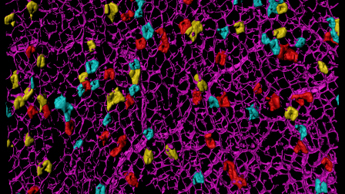
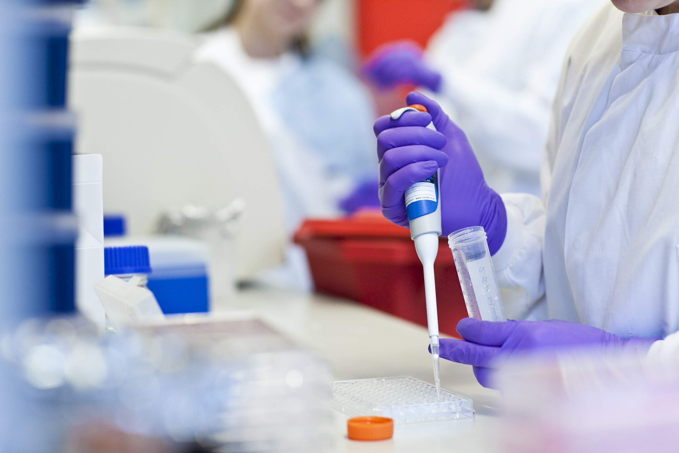
Microscopy
Providing expertise, training and instrumentation for a range of advanced light microscopy techniques across scales and for a variety of samples.
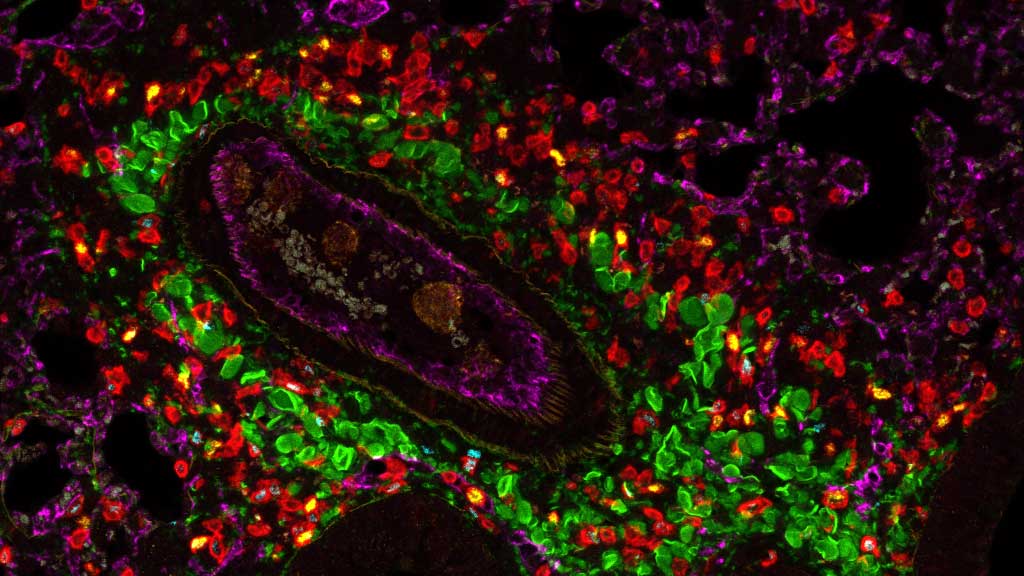
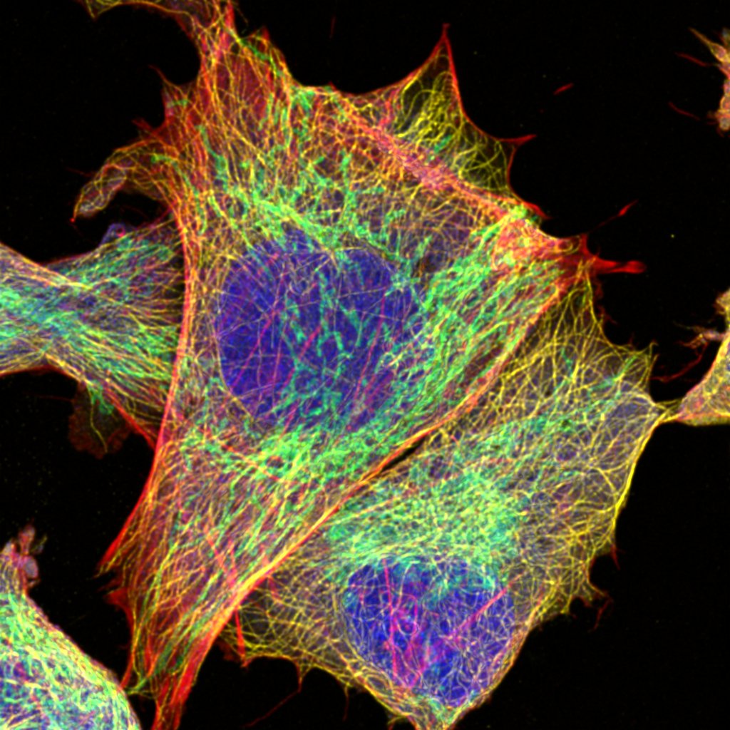
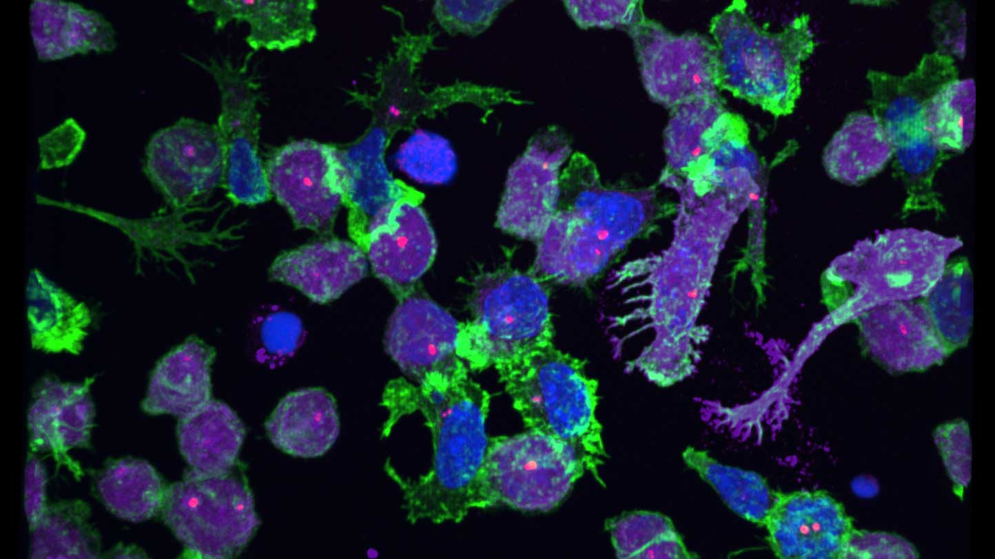
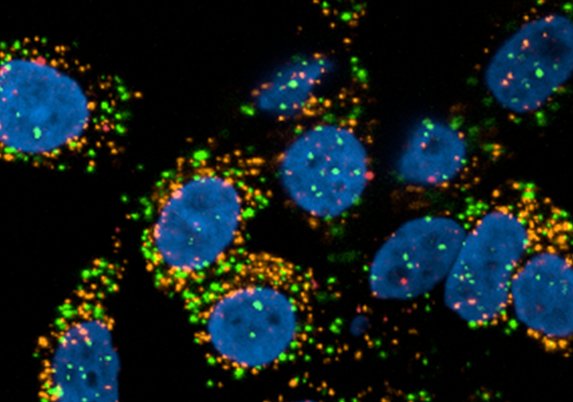
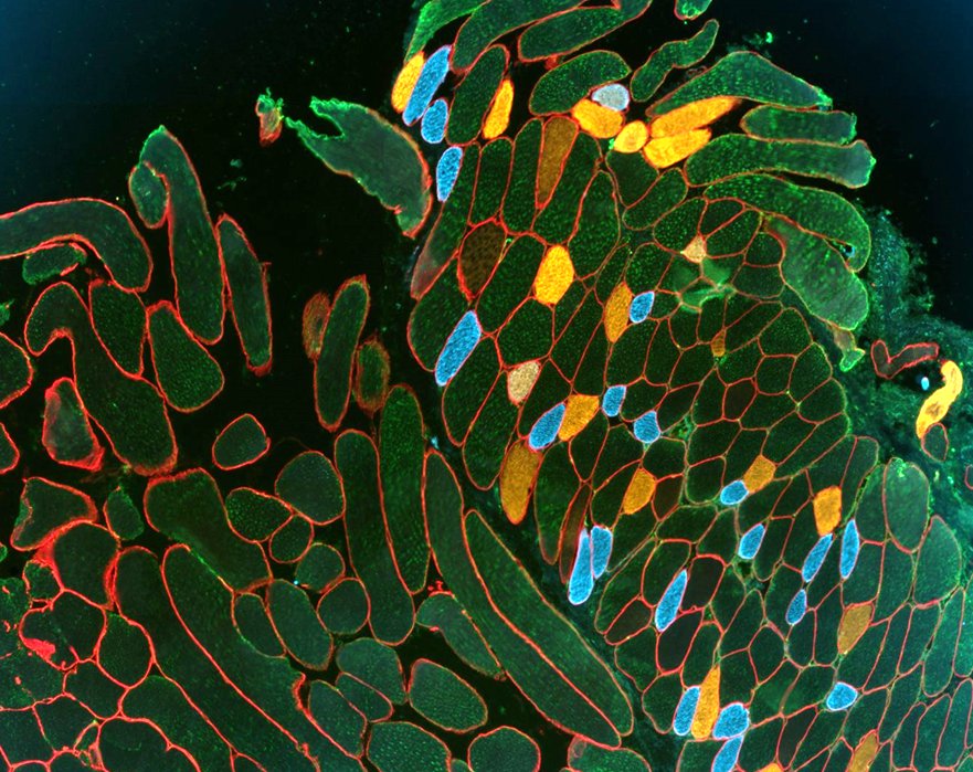
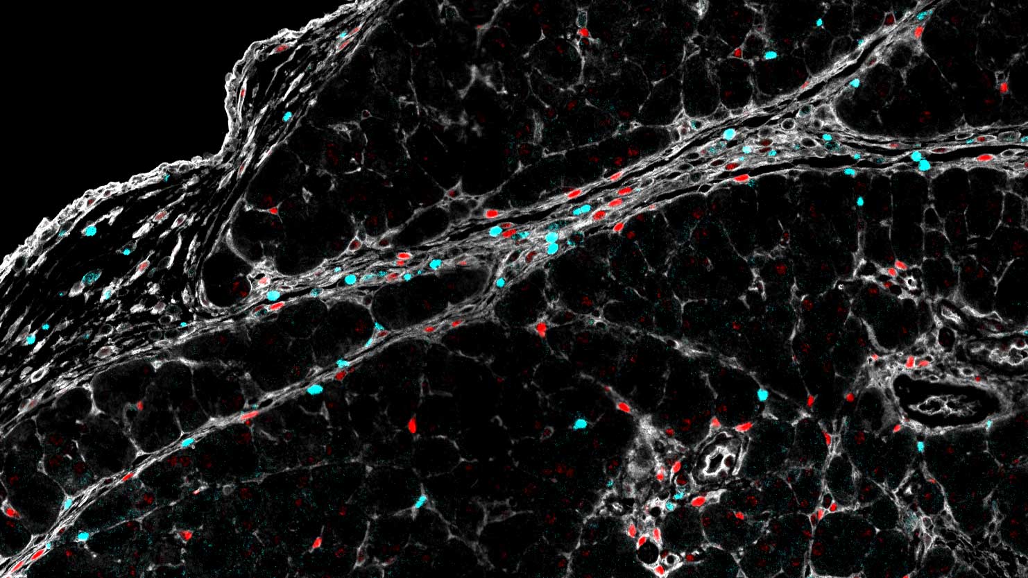
We provide training and support to scientists in a wide range of image acquisition and analysis techniques. Instruments in our facility range from wide-field to non-linear multi-photon and lifetime imaging systems for cell and tissue studies. We are specialised in the following areas of expertise:
- Advanced live-cell imaging using wide-field and spinning disc imaging systems
- Laser scanning confocal microscopy
- Quantitative high content image acquisition and analysis
- Light sheet microscopy of living and cleared samples
- STED super-resolution microscopy
- Non-linear imaging techniques such as multi-photon and second harmonic imaging
- Qualitative fluorescence life-time imaging (TauSense)

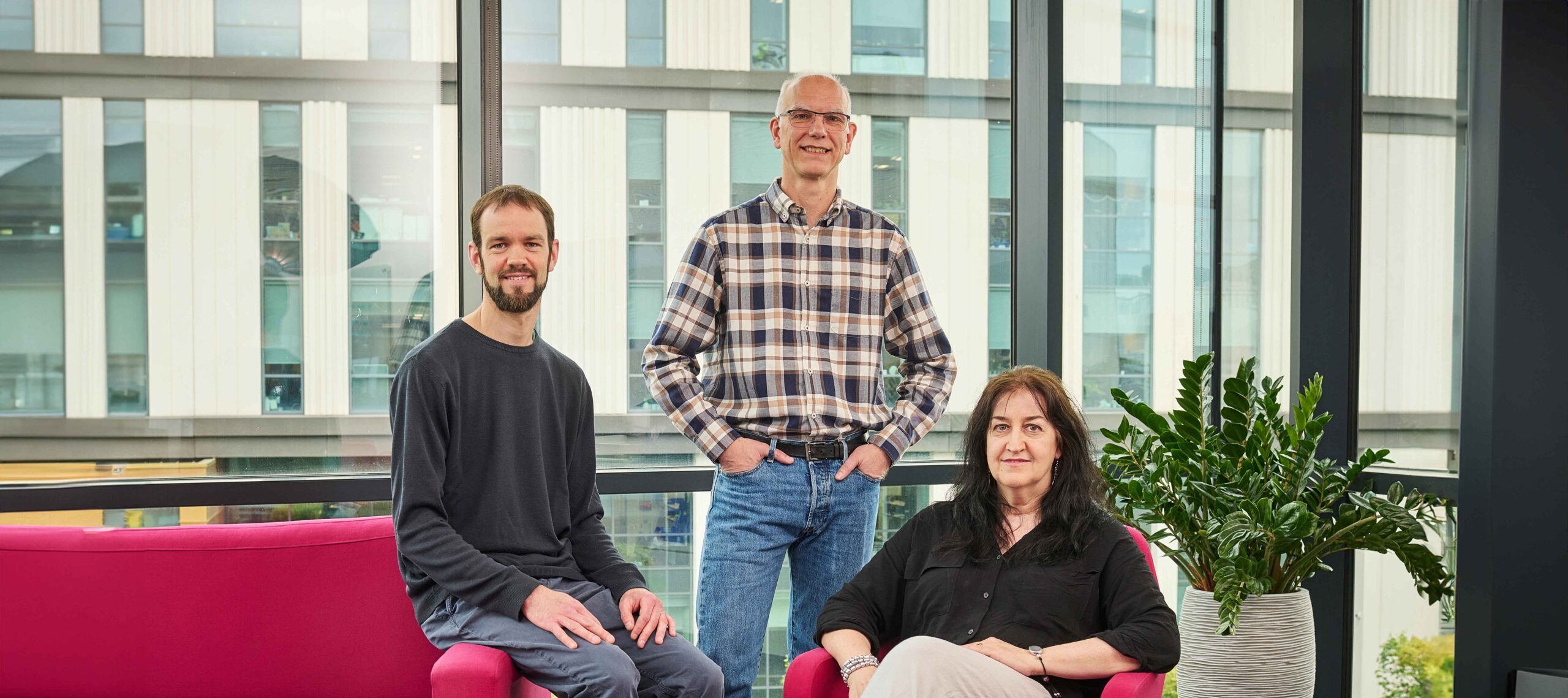
Dr Andreas Bruckbauer
Core Facility Manager
Equipment
Leica Stellaris 8 – MP
Confocal microscope for high resolution and flexible 3D imaging of live and fixed samples.
- Extended white light laser (440 nm – 790 nm) and 405 nm laser
- New sensitive Hybrid detectors including one with far-red sensitivity, photon counting mode (3x HyDS, 1x HyDX, 1x HyDR)
- Lightning deconvolution
- TauSense qualitative lifetime imaging
- Two-Photon imaging for collagen and deep tissue fluorescence imaging with 680 to 1080 nm excitation
- 8000 Hz resonance scanner
- AOBS (acoustical optical beam splitter)
- Navigator and Dye Assistant
- Spectral detection (excitation and emission scans)
- High resolution and glycerol objectives
- Transmitted light DIC (40x, 63x, 100x)
- Stage incubator for live cell imaging
Leica SP8 – STED
Confocal microscope for high resolution and flexible 3D imaging of live and fixed samples with super-resolution capabilities.
- White light laser 470 nm – 670 nm
- 405 and 442 nm lasers
- 5 detectors, 2 PMTs and 3 sensitive HyD detectors, gated
- 8000 Hz resonance scanner
- AOBS (acoustical optical beam splitter)
- Deconvolution with Huygens software
- STED super-resolution imaging with 594. 660, and pulsed 775 nm depletion lasers
- Spectral detection (excitation and emission scans)
- Transmitted light DIC
- Navigator and Dye assistant
- Incubation for live cell imaging with CO2 control
- Cooling stage
Dragonfly Spinning Disk
Laser-based Spinning Disk Confocal microscope for fast 3D imaging of fixed and live samples.
- Lasers: 405, 445, 488, 561, 637 and 730 nm
- two EMCCD cameras Andor iXon 888
- Stage top incubator with CO2 and humidity control.

Opera Phenix Plus
Laser-based spinning disc confocal system for high content cell screening and profiling, volumetric imaging of organoids, spheroids and live cell timelapse imaging.
- Five laser lines at 375 nm, 425 nm, 488 nm, 561 nm and 640 nm.
- Four cameras offering fast acquisition times.
- Synchrony optics with enhanced separation of multiple fluorescence channels.
- Full environmental control including temperature, CO2 and humidity for long term imaging..
- Automated plate handling robotics and incubator for high throughput imaging of plates or slides and long-time interval imaging of organoids and live cells.
Operetta CLS
LED-based Widefield and Spinning Disc Confocal system for high content cell screening and profiling, volumetric imaging of organoids, spheroids, and live cell timelapse imaging.
- Eight LEDs at 365 nm, 405 nm, 440 nm, 475 nm. 510 nm, 550 nm, 630 nm and 660 nm.
- Ideal for highly multiplexed immunofluorescence imaging up to 6 colours.
- Environmental control including temperature and CO2 for long term imaging.
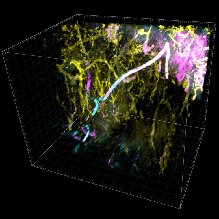
Zeiss Light Sheet Z1
Fast, gentle multiview imaging of large specimens using Light Sheet Microscopy.
- Lasers: 405, 488, 561 and 638 nm
- 2 cameras pco.edge 4.2 for simultaneous imaging.
- incubation chamber with CO2 control for live imaging.
- Cleared tissue with refractive index of 1.58
Zeiss Widefield 1&2
Widefield imaging with transmitted light brightfield, DIC and reflected light fluorescence with LED excitation.
- Fully motorized Axio-Observer Z.1
- Definite Focus
- Incubation chamber with CO2 control.
System 1: Fluorescent proteins, fast dynamics
- Camera: Hamamatsu Orca Falsh 4
- Light source: Lumencore Spectra X
- Filter cubes: CFP, YFP, multi-band: DAPI, FITC, Rhod, DIC
System 2: Standard dyes + NIR fluorescence
- Camera: AxioCam 506
- Light sources: CoolLED pE 4000 and pE-2
- Filter cubes: DAPI, GFP, DsRed, RFP, Cy5, Cy7, and DIC
Software
We provide access to the following commercial image analysis software on our high-performance workstations or remote access servers.
- Harmony (Revvity, formerly PerkinElmer): High-Content imaging analysis software for Opera Phenix and Operetta CLS microscopes
- Vision 4D (Zeiss, formerly Arivis): Image analysis for 2D, 3D and 4D data sets, ideal for Light Sheet microscopy data
- Imaris (Oxford Instruments, formerly Bitplane): 3D and 4D data sets
- Huygens (Scientific Volume Imaging): Deconvolution of 3D data sets
- VitroVivo (Revvity Signals Research Suite): Data processing and dimensionality reduction


-
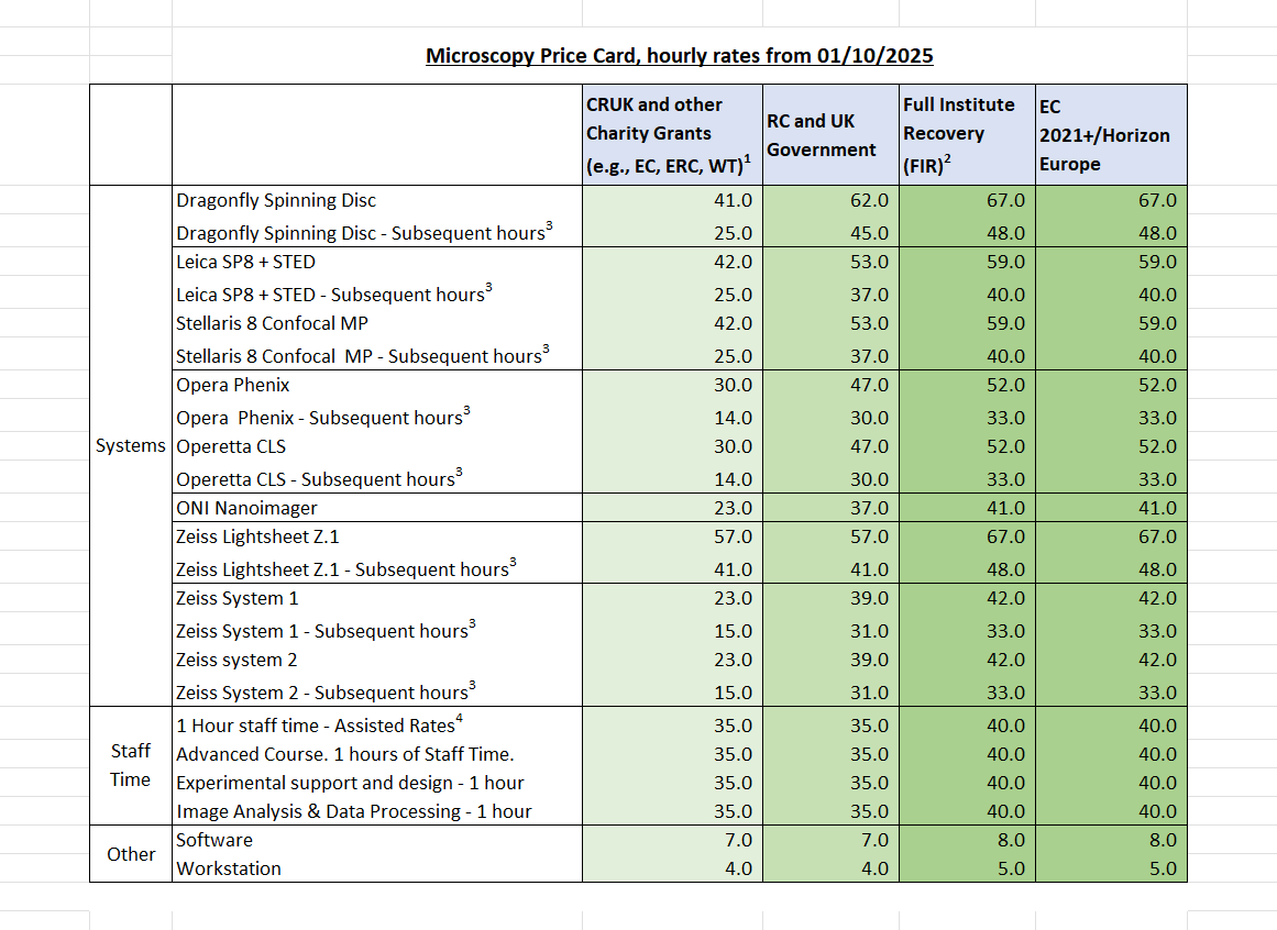
Costs and charging
The institute recovers costs from the grants and projects used in the core facilities and this is reviewed regularly. The hourly rates (in £) for the instruments and services within the core can be found here.
Related News
See all news-
New immune pathway offers treatment hope for childhood brain tumours
3rd February 2026
A newly discovered immune pathway could lead to gentler treatments for multiple childhood brain cancers, according to new research from our Gilbertson Group published today in Nature Genetics.
Find out more -
Order of cancer-driving mutations affects the chance of tumour development
3rd December 2025
New research from the Winton Group has revealed that the order of cancer-driving mutations plays an important role in whether tumours in the intestine can develop.
Find out more -
Single-cell study sheds new light on why ovarian cancer becomes resistant to chemotherapy
11th August 2025
Researchers at the Cancer Research UK Cambridge Institute and Stanford University have mapped how ovarian cancer cells respond to chemotherapy at an unprecedented level of detail, offering new insights into why treatment resistance develops.
Find out more
Laboratory Efficiency Assessment Framework (LEAF)
Microscopy contributed to the Institute’s LEAF Silver accreditation, see the Sustainability webpage for more information.