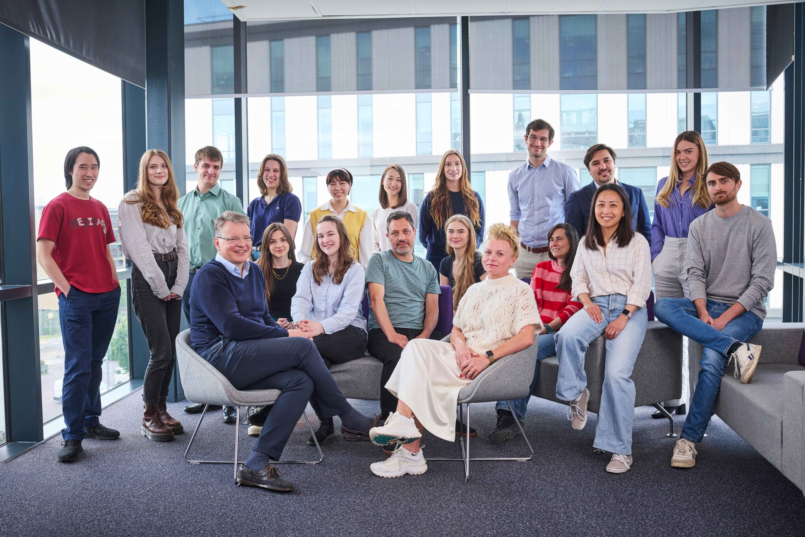
Brenton Group
Functional genomics of ovarian cancer
Research summary
We study the genetic changes that happen in ovarian cancer to understand what causes cancer cells to become resistant to treatment. We use medical imaging technology to study how tumours respond to treatment and also to help us study cancers that have spread to other parts of the body. By understanding how drug resistance develops, we hope to improve treatments by tailoring drug regimes according to the genetic ‘signature’ of a patient’s cancer.
Introduction
Our laboratory focuses on discovering improved treatments for epithelial ovarian cancer using laboratory and clinical studies.

Professor James Brenton
Senior Group Leader
Research topics
Building a molecular classifier
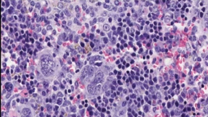
How many types of high grade serous ovarian cancer are there?
The key genomic features of high grade serous ovarian carcinoma (HGSOC) are ubiquitous TP53 mutation and profound genomic instability causing highly abnormal copy number (CN) and structural variation (SV). We have exploited these to develop potential molecular classifications of high-grade serous ovarian cancer (HGSOC).
We have optimised our low-cost tagged amplicon sequencing of TP53, resulting in more accurate mutation calling and variant allele fraction estimation for FFPE and cell free DNA. We have used these assays to validate the performance of p53 IHC as a predictor of TP53 mutation and establish sensitivity and specificity parameters. Our methods have direct clinical impact and have been incorporated as exploratory endpoints in clinical trials. In addition, we are translating these assays and analysis pipelines into the clinic for use in women with HGSOC.
Recently, we have adopted a low-cost, low-coverage, whole-genome sequencing approach to interrogate the copy-number landscape of HGSOC in a clinical setting. We have developed a bioinformatic method that estimates tumour purity and ploidy from shallow WGS using TP53 mutant allele fraction in order to get absolute copy-number. Application of this to a cohort of 300 samples revealed distinct copy-number signatures.
Looking further forward we plan to leverage the observation made by others that subtypes of HGSOC have immune related gene expression signatures – we will overlay matched H&E image analysis, quantifying immune cell infiltration, with our tagged amplicon and copy-number sequencing to attain a more comprehensive classification.
Unlocking the clinical utility of ctDNA
Circulating tumour DNA is a promising non-invasive biomarker that can provide highly specific genomic information in women with HGSOC.
We have demonstrated that ctDNA TP53 mutant allele fraction is correlated with tumour volume and that a decrease after cycle 1 is associated with longer time to progression. It appears that ctDNA is promising as a predictive and prognostic biomarker in a relapsed setting and we are now validating these findings in further cohorts from other clinical trials.
ctDNA has the potential to provide biomarkers of high clinical utility for cancer diagnosis and monitoring in HGSOC. We will transfer our optimized ctDNA assays into the clinic for implementation in wider clinical trials.
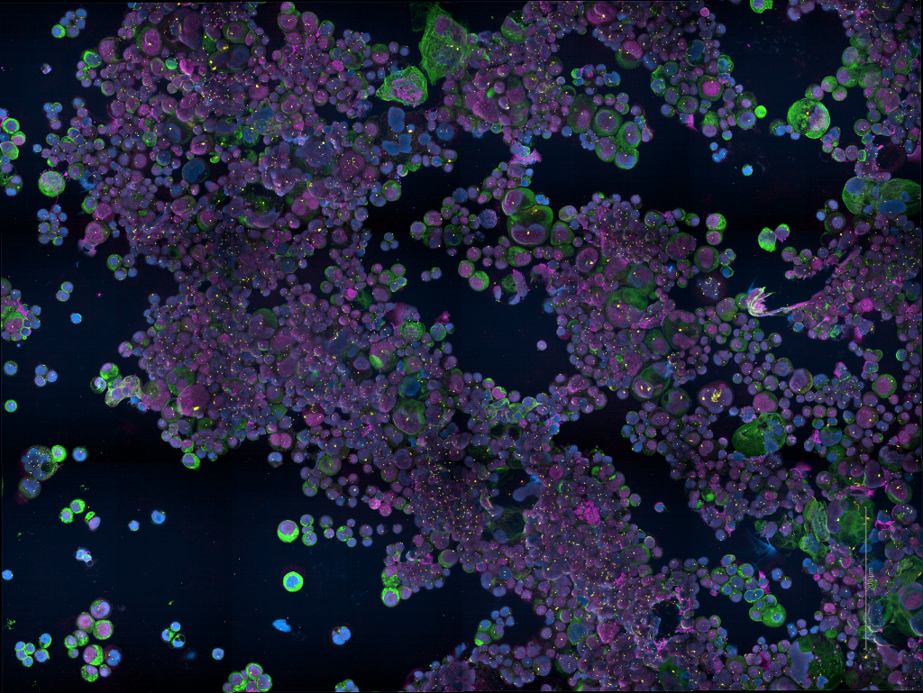
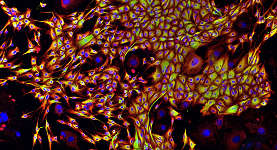
Functional analysis of cellular heterogeneity
In vitro and in vivo tumour models to study HGSOC are very limited. Our aim is to establish reliable pre-clinical model systems that can represent the complex genomic profiles and intratumoural heterogeneity of HGSOC so reagents can be used in functional screens and the discovery of therapeutics and predictive biomarkers.
We have carried out systematic culture experiments to develop 2D cell lines which show that primary HGSOC cells have a highly reproducible limited lifespan when grown as in vitro. We have optimized conditions to grow primary ascites cells in suspension and have tested with RNAseq the similarity between primary ascites spheroids and established cell lines grown in 3D using low attachment media. We have started to develop methods for organoids grown from primary cells and demonstrate improved survival compared to 2D methods.
We are developing established HGSOC xenografts for long term maintenance of HGSOC populations and have optimized methods for tumour disaggregation, freezing, and transplantation. To investigate the cellular heterogeneity within HGSOC, we have used a combination of multi-parameter flow cytometry and in vitro and in vivo functional assays to interrogate the growth and differentiation properties of phenotypically distinct cell populations present within freshly isolated, non-cultured HGSOC tissues.
We have established proof-of-principle methods for characterising and screening patient-derived ascites spheroids in high-throughput assays and for the establishment and functional interrogation of PDX xenograft models.
Related News
See all news-
1M to advance AI powered personalised ovarian cancer care
19th February 2026
Researchers from our Brenton Group are part of an international team awarded the Global Ovarian Cancer Research Consortium’s inaugural AI Accelerator Grant.
Find out more -
Targeting paused cells could improve chemotherapy for lung and ovarian cancers
3rd February 2026
New research published today in Nature Aging by scientists at the University of Cambridge sheds light on why some lung and ovarian cancers stop responding to chemotherapy, and how this resistance might one day be prevented.
Find out more -

Single-cell study sheds new light on why ovarian cancer becomes resistant to chemotherapy
11th August 2025
Researchers at the Cancer Research UK Cambridge Institute and Stanford University have mapped how ovarian cancer cells respond to chemotherapy at an unprecedented level of detail, offering new insights into why treatment resistance develops.
Find out more


Publications
See All PublicationsLaboratory Efficiency Assessment Framework (LEAF)
The Brenton Group contributed to the Institute’s LEAF Silver accreditation, see the Sustainability webpage for more information.