Bohndiek Group
Imaging oxygen and oxidative stress
Research summary
Our research focuses on creating advanced optical devices and systems to improve medical imaging. In doing so, we aim to enhance early cancer detection and understand how blood vessel function impacts tumour evolution.
Introduction
Our core line of research is the creation of innovative optical devices, systems and analysis methods.
We are motivated to answer biomedical research questions, such as understanding how vascular function impacts tumour evolution, and address unmet clinical needs, such as reducing the miss rate for early cancer in endoscopy.

Professor Sarah Bohndiek
Senior Group Leader
Research areas
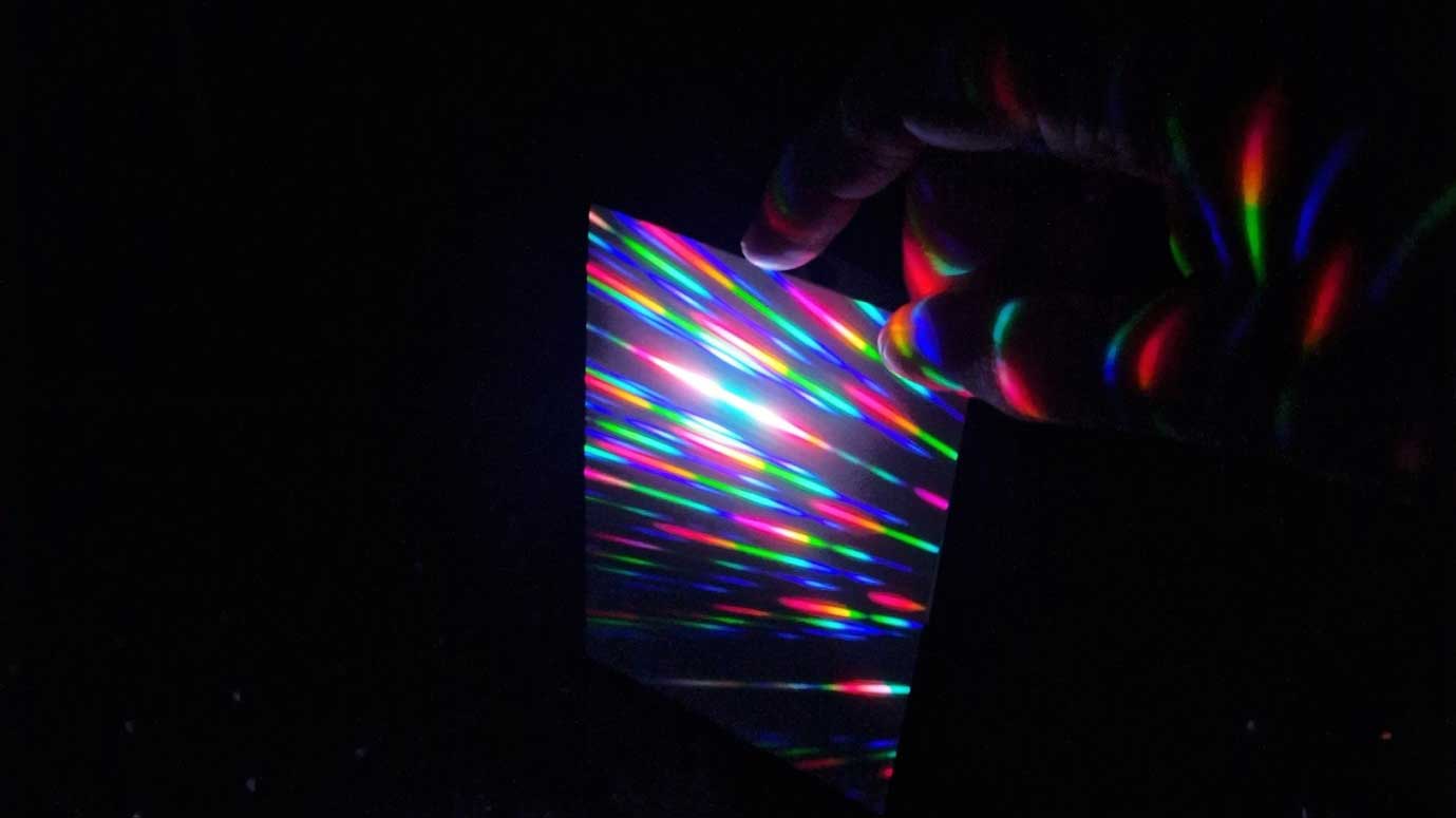
Enabling optical interrogation of complex biological environments
Making measurements at a range of depths in a living breathing patient in a noisy, bright, busy clinical environment presents a profoundly different set of constraints to those commonly found in optics laboratories. For hyperspectral imaging this presents a major challenge, since any distortions or artifacts in the recorded 3D ‘hypercube’ (x,y,wavelength) will lead to misclassification of tissue types.
We have spearheaded efforts to address several of the main factors leading to such distortions, namely motion, miniaturization, and calibration, through advances in physics, chemistry, and data science. By introducing new high-speed line-scan and custom snapshot hyperspectral systems while applying deep-learning analyses we were able to build distortion-free hypercubes – even for handheld operation – and perform real-time classification. We have conceived new optical filter fabrication methods and endoscope designs to miniaturize hyperspectral imaging systems and enable imaging through hair-thin optical fibers.
Importantly for clinical translation, we have led international efforts in standardisation: establishing criteria for biophotonic standards; developing new materials for stable and dynamic tissue-mimicking test objects; and leading global multi-centre evaluations.
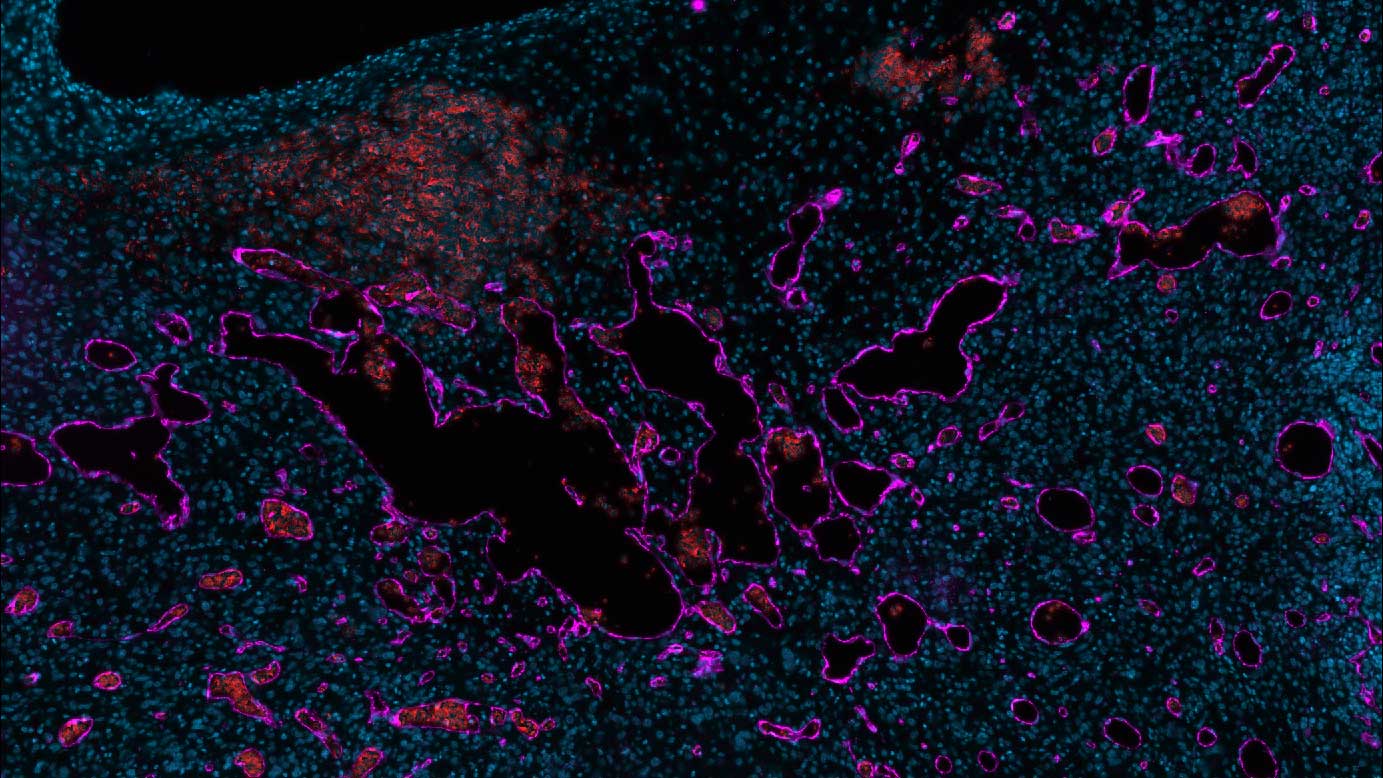
Deriving meaning from imaging measurements to observe biological phenomena
In a clinical context, the interpretation of an image leads to decision-making that impacts patient management. The simplest and easiest optical interaction to measure in humans is the absorption of light by the hugely abundant molecule haemoglobin, which gives red blood cells their colour and allows us to interrogate vascular architecture and function using optics. Equipped with new imaging tools, we are creating a deeper understanding of vascular alterations associated with early pre-cancerous changes and how tumours create and modify their vascular structures to derive oxygen and nutrients during early cancer and in response to therapy. Most recently, we have begun to parameterise these vascular measurements, using both biophysical modelling and machine learning methods, to predict how changes in vessel size and topology contribute to the observable macroscopic effects of dynamic hypoxia exposure.
Cumulatively, our studies, in both cancer models and patients, have caught the attention of the academic community and the public, receiving widespread international media attention.
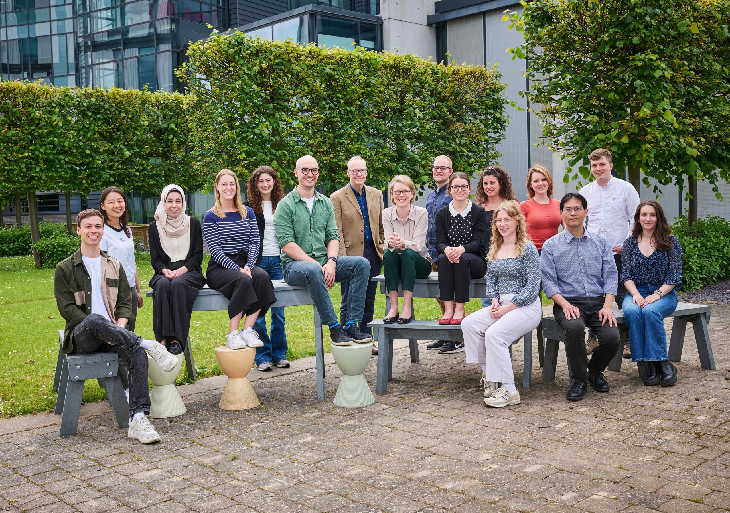
Group Members
-

Chuck Wang
Research Associate
-
Paul Sweeney
Senior Research Associate
-

Janek Grohl
Temporary Staff
-

Ellie Bunce
Postgraduate Student
-
Isabelle Racicot
Research Associate
-

Katie-Lou White
Postgraduate Student
-

Ran Tao
Postgraduate Student
-
Stephen Mead
-
Lorna Wright
Scientific Associate
-
Mariam-Eleni Oraiopoulou
Senior Research Associate
-
Melissa Watt
Postgraduate Student
-

Molly Bridger
Research Administrator
-
Monika Golinska
Senior Scientific Associate
-
Sabrina Bentouati
Research Assistant
-

Seema Bachoo
Postgraduate Student
-

Thierry Lefebvre
Postgraduate Student
-
Thomas Else
Research Associate
Related News
See all news-

Prof Sarah Bohndiek appointed as a Founding Programme Director of ARIA
11th September 2023
Prof Sarah Bohndiek will be working with the Advanced Research and Invention Agency (ARIA), governmental research and development funding agency.
Find out more -
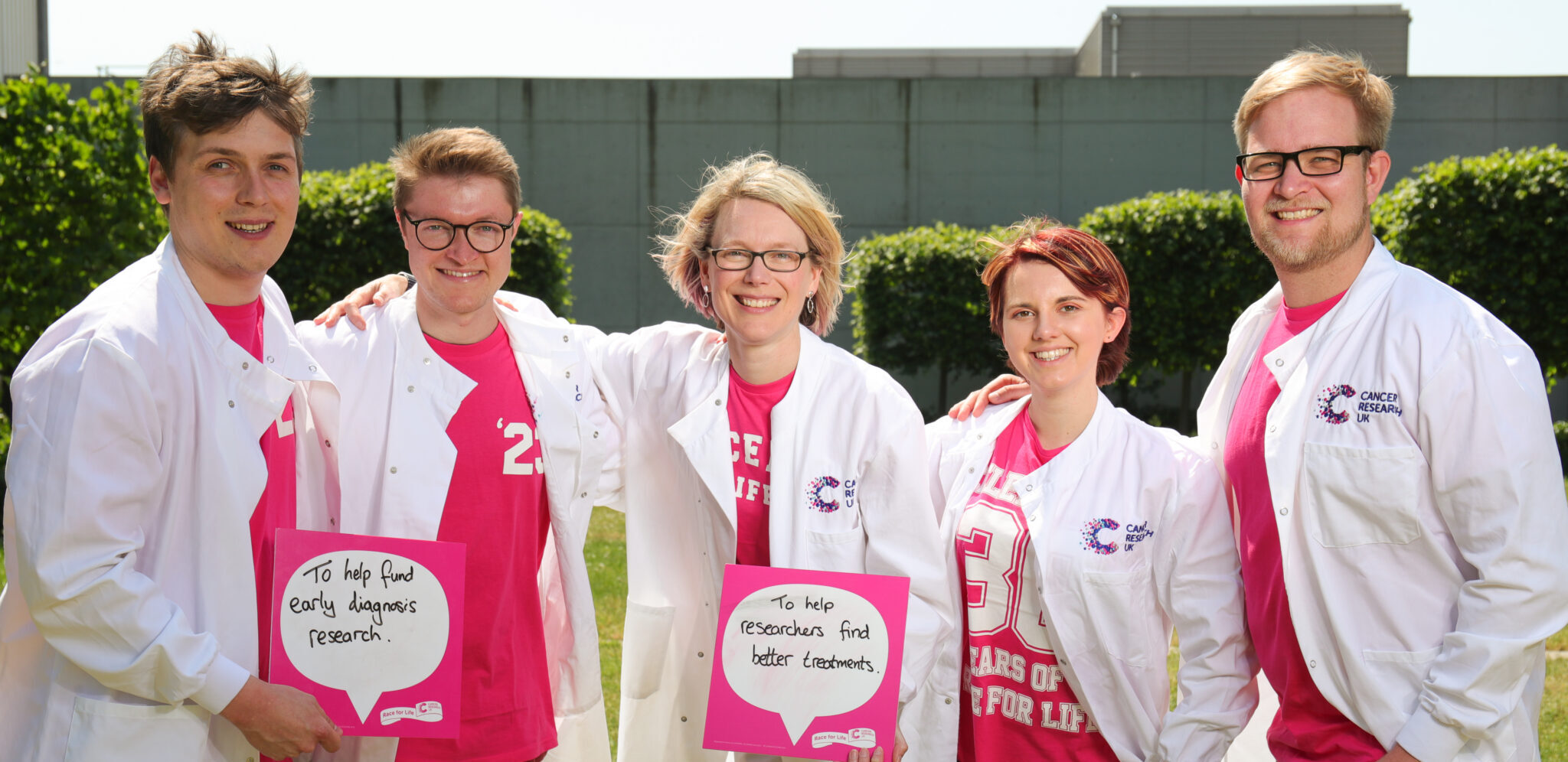
Bohndiek Group to Race for Life
15th June 2023
The Bohndiek Group will be racing alongside Cancer Research UK supporters at this year’s Race for Life 10k at Jesus Green.
Find out more -
Lina Hacker awarded Postgraduate Student Thesis Prize
19th October 2022
The Prize is awarded each year to an exceptional postgraduate student who has completed an outstanding piece of work during their studies.
Find out more
Publications
-
The in vitro dynamics of pseudo-vascular network formation.
E-pub date: 31 Aug 2024
-
Unsupervised Segmentation of 3D Microvascular Photoacoustic Images Using Deep Generative Learning.
E-pub date: 31 Aug 2024
-
Tutorial on phantoms for photoacoustic imaging applications.
E-pub date: 31 Aug 2024
-
Raman micro-spectroscopy reveals the spatial distribution of fumarate in cells and tissues
E-pub date: 4 Jul 2023
Laboratory Efficiency Assessment Framework (LEAF)
The Bohndiek Group contributed to the Institute’s LEAF Silver accreditation, see the Sustainability webpage for more information.